CaMKII delta抗体


Non-transfected (–) and transfected (+) 293T whole cell extracts (50 μg) were separated by 10% SDS-PAGE, and the membrane was blotted with CaMKII delta antibody (GTX111401) diluted at 1:10000. The HRP-conjugated anti-rabbit IgG antibody (GTX213110-01) was used to detect the primary antibody.
Various whole cell extracts (30 μg) were separated by 10% SDS-PAGE, and the membrane was blotted with CaMKII delta antibody (GTX111401) diluted at 1:1000. The HRP-conjugated anti-rabbit IgG antibody (GTX213110-01) was used to detect the primary antibody.

CaMKII delta antibody detects CaMKII delta protein at cytoplasm in rat heart by immunohistochemical analysis.
Sample: Paraffin-embedded rat heart.
CaMKII delta antibody (GTX111401) diluted at 1:400.
Antigen Retrieval: Citrate buffer, pH 6.0, 15 min

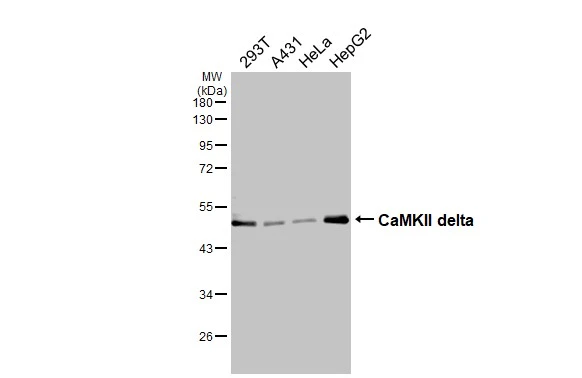
Various tissue extracts (50 μg) were separated by 10% SDS-PAGE, and the membrane was blotted with CaMKII delta antibody (GTX111401) diluted at 1:50000. The HRP-conjugated anti-rabbit IgG antibody (GTX213110-01) was used to detect the primary antibody.
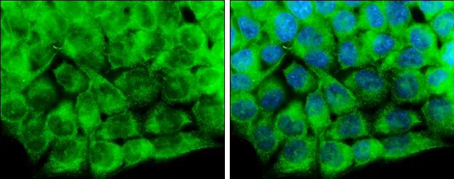
CaMKII delta antibody detects CaMKII delta protein at cytoplasm by immunofluorescent analysis.
Sample: HCT116 cells were fixed in 4% paraformaldehyde at RT for 15 min.
Green: CaMKII delta protein stained by CaMKII delta antibody (GTX111401) diluted at 1:500.
Blue: Hoechst 33342 staining.

CaMKII delta antibody detects CaMKII delta protein at cytoplasm in mouse muscle by immunohistochemical analysis.
Sample: Paraffin-embedded mouse muscle.
CaMKII delta antibody (GTX111401) diluted at 1:400.
Antigen Retrieval: Citrate buffer, pH 6.0, 15 min
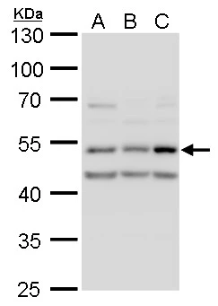
CaMK2D antibody detects CaMK2D protein by western blot analysis.
A. 30 μg A549 whole cell lysate/extract
B. 30 μg H1299 whole cell lysate/extract
C. 30 μg HCT116 whole cell lysate/extract
10% SDS-PAGE
CaMK2D antibody (GTX111401) dilution: 1:1000
The HRP-conjugated anti-rabbit IgG antibody (GTX213110-01) was used to detect the primary antibody.
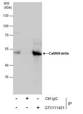
Immunoprecipitation of CaMKII delta protein from HepG2 whole cell extracts using 5 μg of CaMKII delta antibody (GTX111401).
Western blot analysis was performed using CaMKII delta antibody (GTX111401).
EasyBlot anti-Rabbit IgG (GTX221666-01) was used as a secondary reagent.

Zebrafish tissue extract (30 μg) was separated by 10% SDS-PAGE, and the membrane was blotted with CaMKII delta antibody (GTX111401) diluted at 1:40000.
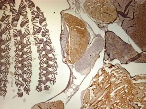
Immunohistochemical analysis of paraffin-embedded zebrafish tissue, using CaMKII delta antibody (GTX111401) at 1:300 dilution.
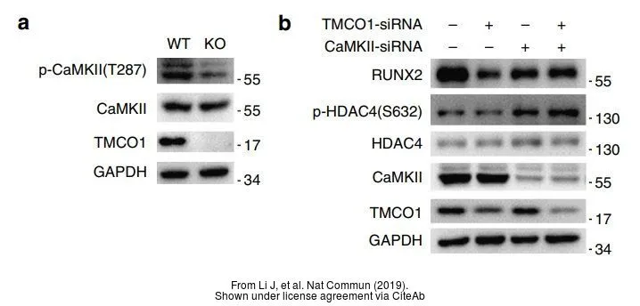
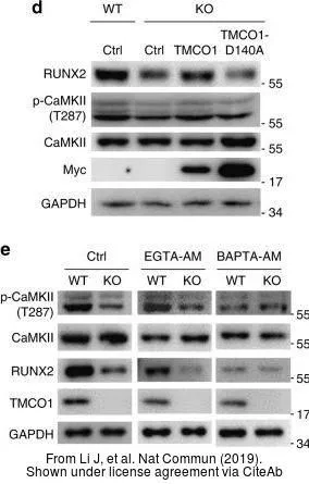
The data was published in the journal Nat Commun in 2019. PMID: 30962442
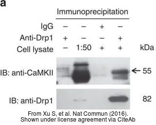
The data was published in the journal Nat Commun in 2016.PMID: 27739424
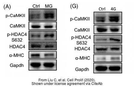
The data was published in the journal Cell Prolif in 2020.PMID: 32101357
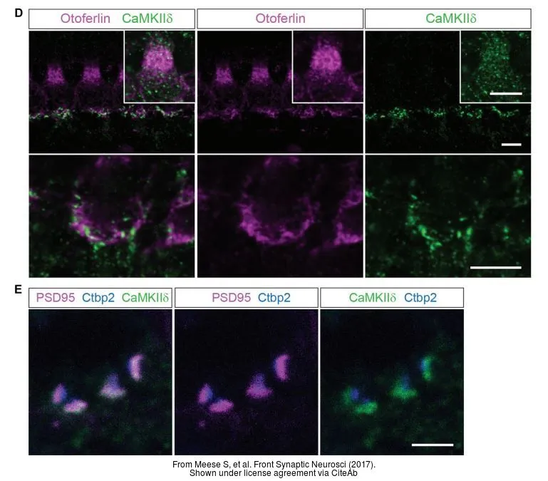
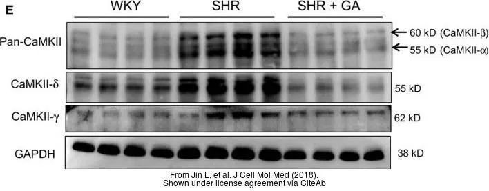
The data was published in the journal Front Synaptic Neurosci in 2017.PMID: 29046633
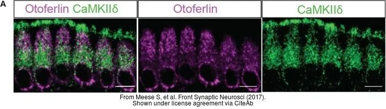
The data was published in the journal Front Synaptic Neurosci in 2017.PMID: 29046633
-
宿主Rabbit
-
克隆Polyclonal
-
同种型IgG
-
实验应用WB ICC/IF IHC-P IP
-
种属反应Human, Mouse, Rat, Zebrafish, Rabbit