Caspase 3抗体


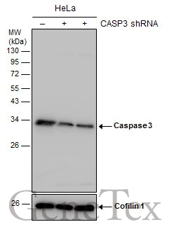
Non-transfected (–) and transfected (+) HeLa whole cell extracts (30 μg) were separated by 12% SDS-PAGE, and the membrane was blotted with Caspase 3 antibody (GTX110543) diluted at 1:4000. The HRP-conjugated anti-rabbit IgG antibody (GTX213110-01) was used to detect the primary antibody.
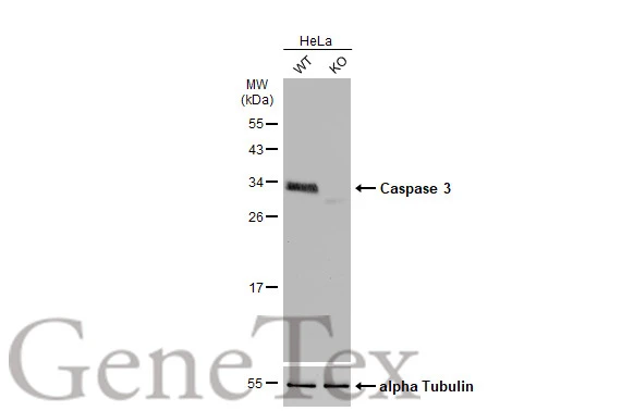
Wild-type (WT) and Caspase 3 knockout (KO) HeLa cell extracts (30 μg) were separated by 12% SDS-PAGE, and the membrane was blotted with Caspase 3 antibody (GTX110543) diluted at 1:5000. The HRP-conjugated anti-rabbit IgG antibody (GTX213110-01) was used to detect the primary antibody.

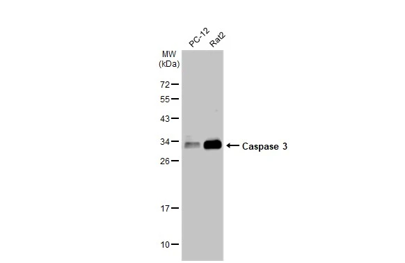
Untreated (–) and treated (+) Rat2 whole cell extracts (30 μg) were separated by 15% SDS-PAGE, and the membrane was blotted with Caspase 3 antibody (GTX110543) diluted at 1:1000. The HRP-conjugated anti-rabbit IgG antibody (GTX213110-01) was used to detect the primary antibody.
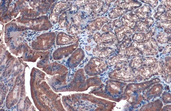
Caspase 3 antibody detects Caspase 3 protein at cytoplasm by immunohistochemical analysis.Sample: Paraffin-embedded mouse duodenum.Caspase 3 stained by Caspase 3 antibody (GTX110543) diluted at 1:1000.Antigen Retrieval: Citrate buffer, pH 6.0, 15 min
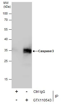
Immunoprecipitation of Caspase 3 protein from HeLa whole cell extracts using 5 μg of Caspase 3 antibody (GTX110543).
Western blot analysis was performed using Caspase 3 antibody (GTX110543).
EasyBlot anti-Rabbit IgG (GTX221666-01) was used as a secondary reagent.
Caspase 3 antibody detects Caspase 3 protein by western blot analysis.
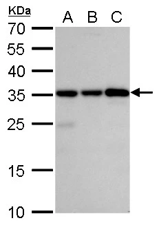
A. 30 μg Jurkat whole cell lysate/extract
B. 30 μg Raji whole cell lysate/extract
C. 30 μg NCI-H929 whole cell lysate/extract
12% SDS-PAGE
Caspase 3 antibody (GTX110543) dilution: 1:5000
The HRP-conjugated anti-rabbit IgG antibody (GTX213110-01) was used to detect the primary antibody.

Caspase 3 antibody detects Caspase 3 protein at cytoplasm in rat liver by immunohistochemical analysis.
Sample: Paraffin-embedded rat liver.
Caspase 3 antibody (GTX110543) diluted at 1:500.
Antigen Retrieval: Citrate buffer, pH 6.0, 15 min
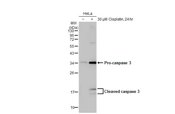
Untreated (–) and treated (+) HeLa whole cell extract (30 μg) were separated by 12% SDS-PAGE, and the membrane was blotted with Caspase 3 antibody (GTX110543) diluted at 1:1000. The HRP-conjugated anti-rabbit IgG antibody (GTX213110-01) was used to detect the primary antibody.
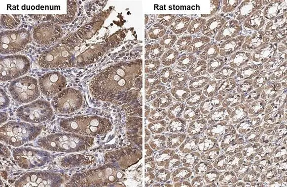
Caspase 3 antibody detects Caspase 3 protein by immunohistochemical analysis.Sample: Paraffin-embedded rat tissues.Caspase 3 stained by Caspase 3 antibody (GTX110543) diluted at 1:500.Antigen Retrieval: Citrate buffer, pH 6.0, 15 min
Various whole cell extracts (30 μg) were separated by 12% SDS-PAGE, and the membrane was blotted with Caspase 3 antibody (GTX110543) diluted at 1:1000. The HRP-conjugated anti-rabbit IgG antibody (GTX213110-01) was used to detect the primary antibody.
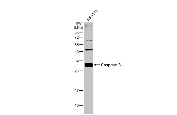
Whole cell extract (30 μg) was separated by 12% SDS-PAGE, and the membrane was blotted with Caspase 3 antibody (GTX110543) diluted at 1:1000. The HRP-conjugated anti-rabbit IgG antibody (GTX213110-01) was used to detect the primary antibody.

Various whole cell extracts (30 μg) were separated by 12% SDS-PAGE, and the membrane was blotted with Caspase 3 antibody (GTX110543) diluted at 1:1000. The HRP-conjugated anti-rabbit IgG antibody (GTX213110-01) was used to detect the primary antibody.
-
宿主Rabbit
-
克隆Polyclonal
-
同种型IgG
-
实验应用WB IHC-P IHC-Fr IP Dot
-
种属反应Human, Mouse, Rat