Caspase 7抗体


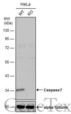
Wild-type (WT) and Caspase 7 knockout (KO) HeLa cell extracts (30 μg) were separated by 10% SDS-PAGE, and the membrane was blotted with Caspase 7 antibody (GTX123679) diluted at 1:1000. The HRP-conjugated anti-rabbit IgG antibody (GTX213110-01) was used to detect the primary antibody.
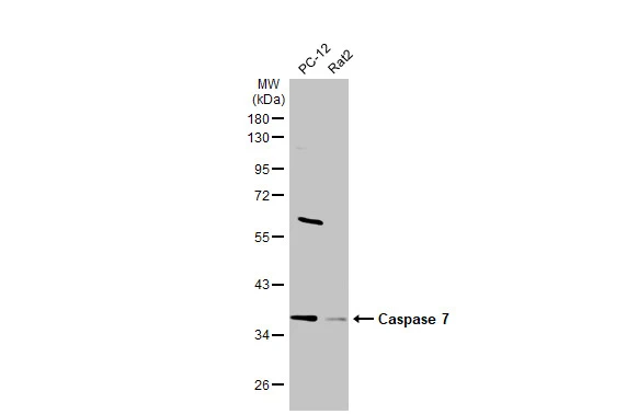
Various whole cell extracts (30 μg) were separated by 10% SDS-PAGE, and the membrane was blotted with Caspase 7 antibody (GTX123679) diluted at 1:1000. The HRP-conjugated anti-rabbit IgG antibody (GTX213110-01) was used to detect the primary antibody.
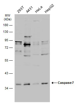
Various whole cell extracts (30 μg) were separated by 10% SDS-PAGE, and the membrane was blotted with Caspase 7 antibody (GTX123679) diluted at 1:1000. The HRP-conjugated anti-rabbit IgG antibody (GTX213110-01) was used to detect the primary antibody.
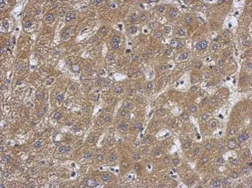
Immunohistochemical analysis of paraffin-embedded human hepatoma, using Caspase 7 p11 subunit(GTX123679) antibody at 1:500 dilution.
Antigen Retrieval: Trilogy™ (EDTA based, pH 8.0) buffer, 15min
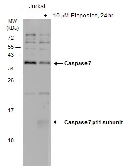
Untreated (–) and treated (+) Jurakt whole cell extracts (30 μg) were separated by 15% SDS-PAGE, and the membrane was blotted with Caspase 7 antibody (GTX123679) diluted at 1:500. The HRP-conjugated anti-rabbit IgG antibody (GTX213110-01) was used to detect the primary antibody, and the signal was developed with Trident ECL plus-Enhanced.
-
宿主Rabbit
-
克隆Polyclonal
-
同种型IgG
-
实验应用WB IHC-P IP
-
种属反应Human, Rat