Caspase 8抗体


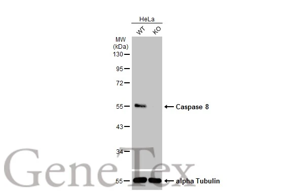
Wild-type (WT) and CASP8 (KO) HeLa cell extracts (30 μg) were separated by 10% SDS-PAGE, and the membrane was blotted with Caspase 8 antibody (GTX110723) diluted at 1:2000. The HRP-conjugated anti-rabbit IgG antibody (GTX213110-01) was used to detect the primary antibody.
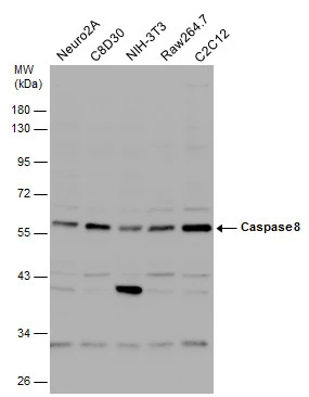
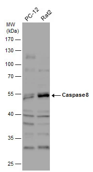
Various whole cell extracts (30 μg) were separated by 10% SDS-PAGE, and the membrane was blotted with Caspase 8 antibody (GTX110723) diluted at 1:1000. The HRP-conjugated anti-rabbit IgG antibody (GTX213110-01) was used to detect the primary antibody.

Various whole cell extracts (30 μg) were separated by 7.5% SDS-PAGE, and the membrane was blotted with Caspase 8 antibody (GTX110723) diluted at 1:1000. The HRP-conjugated anti-rabbit IgG antibody (GTX213110-01) was used to detect the primary antibody. Corresponding RNA expression data for the same cell lines are based on Human Protein Atlas program.
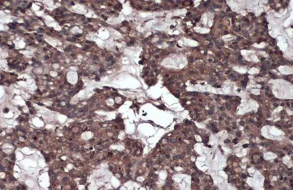
Caspase 8 antibody detects Caspase 8 protein at cytoplasm by immunohistochemical analysis.Sample: Paraffin-embedded human breast carcinoma.Caspase 8 stained by Caspase 8 antibody (GTX110723) diluted at 1:1000.Antigen Retrieval: Citrate buffer, pH 6.0, 15 min
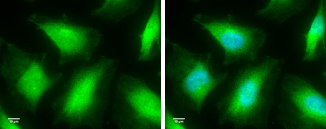
Caspase 8 antibody detects Caspase 8 protein at cytoplasm by immunofluorescent analysis.
Sample: HeLa cells were fixed in 4% paraformaldehyde for 10 min.
Green: Caspase 8 protein stained by Caspase 8 antibody (GTX110723) diluted at 1:100.
Blue: Hoechst 33342 staining.
Scale bar = 10 μm.
Various whole cell extracts (30 μg) were separated by 10% SDS-PAGE, and the membrane was blotted with Caspase 8 antibody (GTX110723) diluted at 1:1000. The HRP-conjugated anti-rabbit IgG antibody (GTX213110-01) was used to detect the primary antibody.
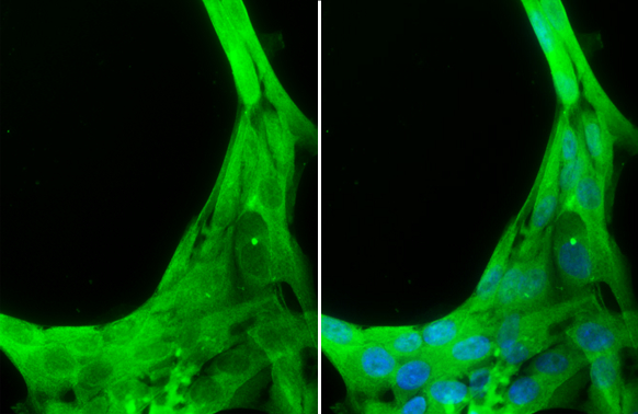
Caspase 8 antibody detects Caspase 8 protein at cytoplasm by immunofluorescent analysis.Sample: PG-4 cells were fixed in 4% paraformaldehyde at RT for 15 min.Green: Caspase 8 stained by Caspase 8 antibody (GTX110723) diluted at 1:100.Blue: Hoechst 33342 staining.
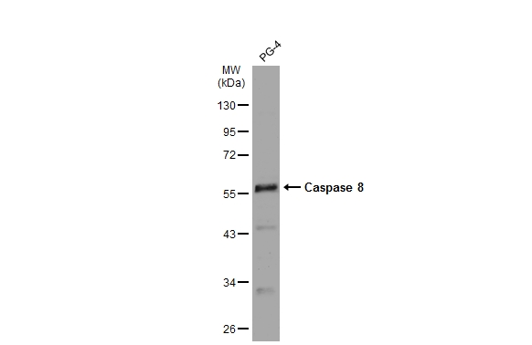
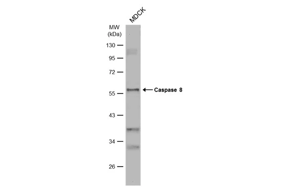
Whole cell extract (30 μg) was separated by 10% SDS-PAGE, and the membrane was blotted with Caspase 8 antibody (GTX110723) diluted at 1:1000. The HRP-conjugated anti-rabbit IgG antibody (GTX213110-01) was used to detect the primary antibody.
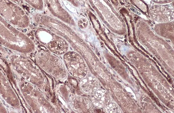
Caspase 8 antibody detects Caspase 8 protein at cytoplasm by immunohistochemical analysis.Sample: Paraffin-embedded cat kidney.Caspase 8 stained by Caspase 8 antibody (GTX110723) diluted at 1:500.Antigen Retrieval: Citrate buffer, pH 6.0, 15 min
Whole cell extract (30 μg) was separated by 10% SDS-PAGE, and the membrane was blotted with Caspase 8 antibody (GTX110723) diluted at 1:1000. The HRP-conjugated anti-rabbit IgG antibody (GTX213110-01) was used to detect the primary antibody, and the signal was developed with Trident ECL plus-Enhanced.

-
宿主Rabbit
-
克隆Polyclonal
-
同种型IgG
-
实验应用WB ICC/IF IHC-P
-
种属反应Human, Mouse, Rat, Cat, Dog