ERp57抗体


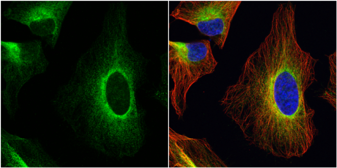
ERp57 antibody detects ERp57 protein at endoplasmic reticulum by immunofluorescent analysis.
Sample: HeLa cells were fixed in 4% paraformaldehyde at RT for 15 min.
Green: ERp57 protein stained by ERp57 antibody (GTX113719) diluted at 1:1000.
Red: alpha Tubulin, a cytoskeleton marker, stained by alpha Tubulin antibody [GT114] (GTX628802) diluted at 1:1000.
Blue: Hoechst 33342 staining.
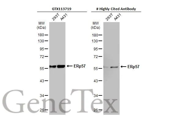
Various whole cell extracts (30 μg) were separated by 10% SDS-PAGE, and the membranes were blotted with ERp57 antibody (GTX113719) diluted at 1:5000 and competitor's antibody diluted at 1:5000. The HRP-conjugated anti-rabbit IgG antibody (GTX213110-01) was used to detect the primary antibody.
*The competitor is not affiliated with GeneTex and does not endorse this product.
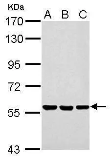
Sample (30 ug of whole cell lysate)
A: Neuro2A
B: GL261
C: C8D30
7.5% SDS PAGE
GTX113719 diluted at 1:1000
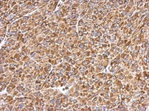
Immunohistochemical analysis of paraffin-embedded U87 xenograft, using ERp57(GTX113719) antibody at 1:500 dilution.
Antigen Retrieval: Trilogy™ (EDTA based, pH 8.0) buffer, 15min

ERp57 antibody detects ERp57 protein at cytoplasm on mouse testis by immunohistochemical analysis.
Sample: Paraffin-embedded mouse testis.
ERp57 antibody (GTX113719) diluted at 1:500.
Antigen Retrieval: Trilogy™ (EDTA based, pH 8.0) buffer, 15min
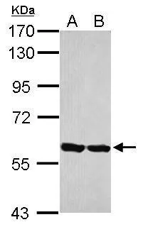
Sample (30 ug of whole cell lysate)
A: HepG2
B: HCT116
7.5% SDS PAGE
GTX113719 diluted at 1:10000
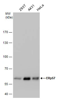
ERp57 antibody detects ERp57 protein by western blot analysis. Various whole cell extracts (30 μg) were separated by 7.5% SDS-PAGE, and the membrane was blotted with ERp57 antibody (GTX113719) diluted by 1:10000.
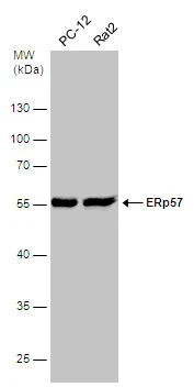
Various whole cell extracts (30 μg) were separated by 10% SDS-PAGE, and the membrane was blotted with ERp57 antibody (GTX113719) diluted at 1:500.
-
宿主Rabbit
-
克隆Polyclonal
-
同种型IgG
-
实验应用WB ICC/IF IHC-P
-
种属反应Human, Mouse, Rat