FAK (phospho Tyr397)抗体


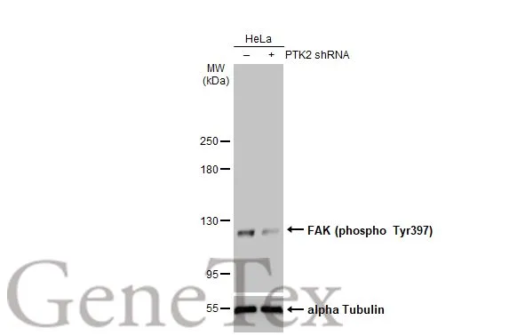
Non-transfected (–) and transfected (+) HeLa whole cell extracts (30 μg) were separated by 5% SDS-PAGE, and the membrane was blotted with FAK (phospho Tyr397) antibody (GTX129840) diluted at 1:500. The HRP-conjugated anti-rabbit IgG antibody (GTX213110-01) was used to detect the primary antibody.

Various whole cell extracts (30 μg) were separated by 7.5% SDS-PAGE, and the membrane was blotted with FAK (phospho Tyr397) antibody (GTX129840) diluted at 1:500. The HRP-conjugated anti-rabbit IgG antibody (GTX213110-01) was used to detect the primary antibody.

Untreated (–) and treated (+) HepG2 whole cell extracts (30 μg) were separated by 7.5% SDS-PAGE, and the membrane was blotted with FAK (phospho Tyr397) antibody (GTX129840) diluted at 1:500. The HRP-conjugated anti-rabbit IgG antibody (GTX213110-01) was used to detect the primary antibody.

Various whole cell extracts (30 μg) were separated by 5% SDS-PAGE, and the membrane was blotted with FAK (phospho Tyr397) antibody (GTX129840) diluted at 1:500. The HRP-conjugated anti-rabbit IgG antibody (GTX213110-01) was used to detect the primary antibody.

Untreated (–) and treated (+) HepG2 whole cell extracts (30 μg) were separated by 5% SDS-PAGE, and the membrane was blotted with FAK (phospho Tyr397) antibody (GTX129840) diluted at 1:500. The HRP-conjugated anti-rabbit IgG antibody (GTX213110-01) was used to detect the primary antibody.
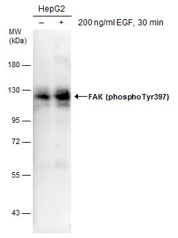
Untreated (–) and treated (+) HepG2 whole cell extracts (30 μg) were separated by 7.5% SDS-PAGE, and the membrane was blotted with FAK (phospho Tyr397) antibody (GTX129840) diluted at 1:500.
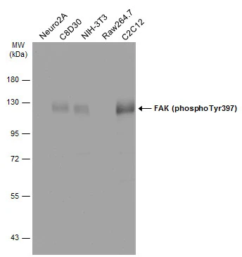
Various whole cell extracts (30 μg) were separated by 7.5% SDS-PAGE, and the membrane was blotted with FAK (phospho Tyr397) antibody (GTX129840) diluted at 1:500. The HRP-conjugated anti-rabbit IgG antibody (GTX213110-01) was used to detect the primary antibody.
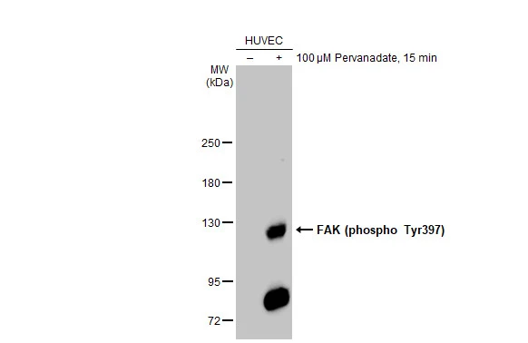
Untreated (–) and treated (+) HUVEC whole cell extracts (30 μg) were separated by 5% SDS-PAGE, and the membrane was blotted with FAK (phospho Tyr397) antibody (GTX129840) diluted at 1:50000. The HRP-conjugated anti-rabbit IgG antibody (GTX213110-01) was used to detect the primary antibody.
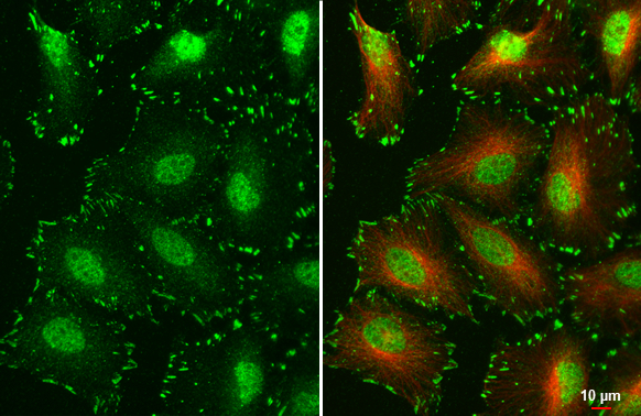
FAK (phospho Tyr397) antibody detects FAK (phospho Tyr397) protein at cell junction, nucleus and cytoplasm by immunofluorescent analysis.Sample: HeLa cells were fixed in 4% paraformaldehyde at RT for 15 min.Green: FAK (phospho Tyr397) stained by FAK (phospho Tyr397) antibody (GTX129840) diluted at 1:500.Red: alpha Tubulin, a cytoskeleton marker, stained by alpha Tubulin antibody [GT114] (GTX628802) diluted at 1:1000.
-
宿主Rabbit
-
克隆Polyclonal
-
同种型IgG
-
实验应用WB ICC/IF IHC-P
-
种属反应Human, Mouse, Rat