FGF13抗体


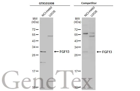
Various whole cell extracts (30 μg) were separated by 12% SDS-PAGE, and the membranes were blotted with FGF13 antibody (GTX101008) diluted at 1:500 and competitor's antibody (Competitor) diluted at 1:500. The HRP-conjugated anti-rabbit IgG antibody (GTX213110-01) was used to detect the primary antibody.
*The competitor is not affiliated with GeneTex and does not endorse this product.

Various whole cell extracts (30 μg) were separated by 12% SDS-PAGE, and the membrane was blotted with FGF13 antibody (GTX101008) diluted at 1:1000. The HRP-conjugated anti-rabbit IgG antibody (GTX213110-01) was used to detect the primary antibody. Corresponding RNA expression data for the same cell lines are based on Human Protein Atlas program.
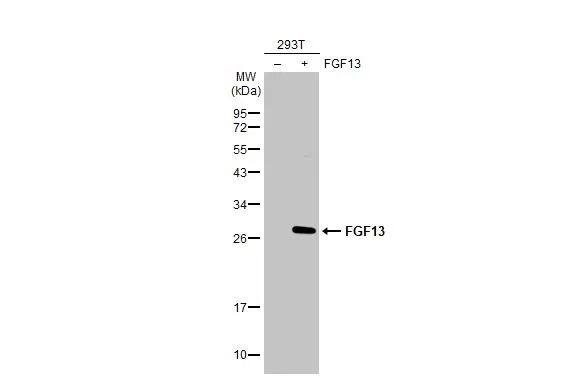
Non-transfected (–) and transfected (+) 293T whole cell extracts (30 μg) were separated by 12% SDS-PAGE, and the membrane was blotted with FGF13 antibody (GTX101008) diluted at 1:5000. The HRP-conjugated anti-rabbit IgG antibody (GTX213110-01) was used to detect the primary antibody.
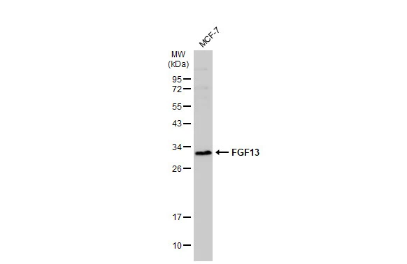
Whole cell extract (30 μg) was separated by 12% SDS-PAGE, and the membrane was blotted with FGF13 antibody (GTX101008) diluted at 1:2000. The HRP-conjugated anti-rabbit IgG antibody (GTX213110-01) was used to detect the primary antibody.
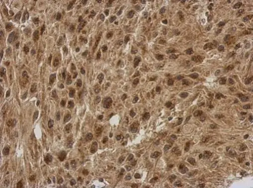
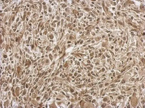
Immunohistochemical analysis of paraffin-embedded U373 xenograft, using FGF13(GTX101008) antibody at 1:500 dilution.
Antigen Retrieval: Trilogy™ (EDTA based, pH 8.0) buffer, 15min
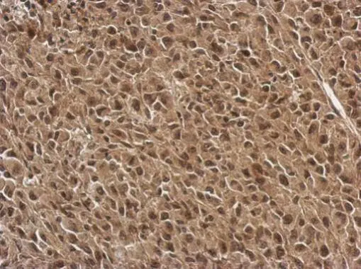
Immunohistochemical analysis of paraffin-embedded C2C12 xenograft, using FGF13(GTX101008) antibody at 1:500 dilution.
Antigen Retrieval: Trilogy™ (EDTA based, pH 8.0) buffer, 15min
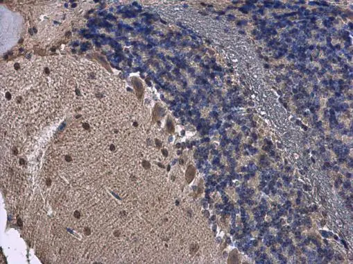
FGF13 antibody detects FGF13 protein at cytoplasm in mouse brain by immunohistochemical analysis.
Sample: Paraffin-embedded mouse brain.
FGF13 antibody (GTX101008) diluted at 1:500.
Antigen Retrieval: Citrate buffer, pH 6.0, 15 min
Immunohistochemical analysis of paraffin-embedded RT2 xenograft, using FGF13(GTX101008) antibody at 1:500 dilution.
Antigen Retrieval: Trilogy™ (EDTA based, pH 8.0) buffer, 15min
-
宿主Rabbit
-
克隆Polyclonal
-
同种型IgG
-
实验应用WB IHC-P
-
种属反应Human, Mouse, Rat