Factor X抗体


HepG2 whole cell extract and conditioned medium (30 μg) were separated by 10% SDS-PAGE, and the membrane was blotted with Factor X antibody (GTX110300) diluted at 1:1000. The HRP-conjugated anti-rabbit IgG antibody (GTX213110-01) was used to detect the primary antibody, and the signal was developed with Trident ECL plus-Enhanced.
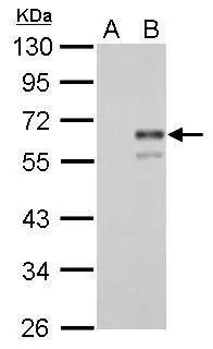
Factor X antibody detects F10 protein by western blot analysis.
A. 30μg 293T whole cell lysate/extract
B. 30 μg whole cell lysate/extract of human F10-transfected 293T cells
10% SDS-PAGE
Factor X antibody (GTX110300) dilution: 1:5000
The HRP-conjugated anti-rabbit IgG antibody (GTX213110-01) was used to detect the primary antibody.
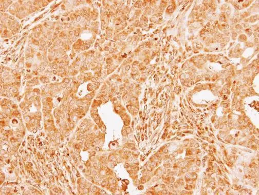
Immunohistochemical analysis of paraffin-embedded NCI-N87 xenograft, using Factor X(GTX110300) antibody at 1:100 dilution.
Antigen Retrieval: Trilogy™ (EDTA based, pH 8.0) buffer, 15min
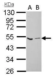
Factor X antibody detects F10 protein by western blot analysis.
A. 30 μg GL261 whole lysate/extract
B. 30 μg C8D30 whole cell lysate/extract
10% SDS-PAGE
Factor X antibody (GTX110300) dilution: 1:1000
The HRP-conjugated anti-rabbit IgG antibody (GTX213110-01) was used to detect the primary antibody.
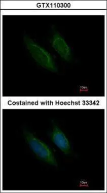
Immunofluorescence analysis of methanol-fixed HeLa, using Factor X(GTX110300) antibody at 1:200 dilution.
-
宿主Rabbit
-
克隆Polyclonal
-
同种型IgG
-
实验应用WB ICC/IF IHC-P
-
种属反应Human, Mouse