Fibrillarin抗体

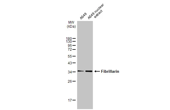
A549 whole cell and nuclear extracts (30 μg) were separated by 12% SDS-PAGE, and the membrane was blotted with Fibrillarin antibody (GTX113684) diluted at 1:500. The HRP-conjugated anti-rabbit IgG antibody (GTX213110-01) was used to detect the primary antibody.
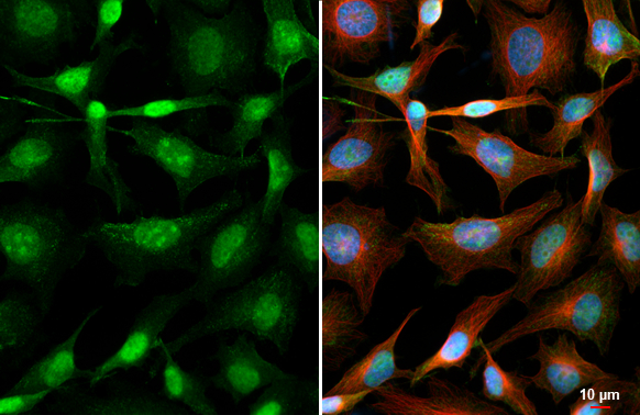
Fibrillarin antibody detects Fibrillarin protein at nucleus, nucleolus and cytoplasm by immunofluorescent analysis.Sample: HeLa cells were fixed in ice-cold MeOH for 5 min.Green: Fibrillarin stained by Fibrillarin antibody (GTX113684) diluted at 1:2000.Red: alpha Tubulin, a cytoskeleton marker, stained by alpha Tubulin antibody [GT114] (GTX628802) diluted at 1:1000.Blue: Fluoroshield with DAPI (GTX30920).

Fibrillarin antibody detects FBL protein by Western blot analysis.
A. 30 μg Neuro2A whole cell lysate/extract
12 % SDS-PAGE
Fibrillarin antibody (GTX113684) dilution: 1:1000
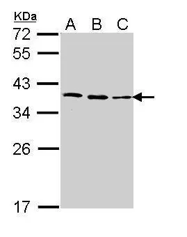
Sample (30 ug of whole cell lysate)
A: 293T
B: H1299
C: Molt-4 (GTX27912)
12% SDS PAGE
GTX113684 diluted at 1:1000
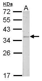
Fibrillarin antibody detects FBL protein by Western blot analysis.
A. 30 μg PC-12 whole cell lysate/extract
12 % SDS-PAGE
Fibrillarin antibody (GTX113684) dilution: 1:500
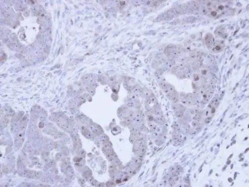
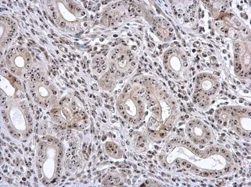
Immunohistochemical analysis of paraffin-embedded NCIN87 Xenograft, using Fibrillarin (GTX113684) antibody at 1:100 dilution.
Antigen Retrieval: Citrate buffer, pH 6.0, 15 min
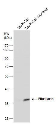
Fibrillarin antibody detects Fibrillarin protein by western blot analysis.
whole cell lysate (30 μg) and nuclear extract (30 μg) from SK-N-SH cells
10 % SDS-PAGE
Fibrillarin antibody (GTX113684) dilution: 1:500

Fibrillarin antibody detects Fibrillarin protein by western blot analysis.
whole cell lysate (30 μg) and nuclear extract (30 μg) from Jurkat cells
10 % SDS-PAGE
Fibrillarin antibody (GTX113684) dilution: 1:500
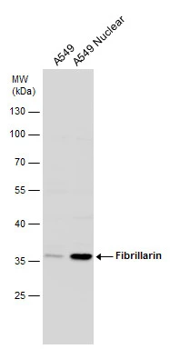
Fibrillarin antibody detects Fibrillarin protein by western blot analysis.
whole cell lysate (30 μg) and nuclear extract(30 μg) from A549 cells
10 % SDS-PAGE
Fibrillarin antibody (GTX113684) dilution: 1:500
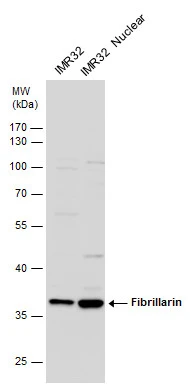
Fibrillarin antibody detects Fibrillarin protein by western blot analysis.
whole cell lysate (30 μg) and nuclear extract (30 μg) from IMR32 cells
10 % SDS-PAGE
Fibrillarin antibody (GTX113684) dilution: 1:500
Fibrillarin antibody detects Fibrillarin protein at nucleolus in mouse cervix by immunohistochemical analysis.
Sample: Paraffin-embedded mouse cervix.
Fibrillarin antibody (GTX113684) diluted at 1:500.
Antigen Retrieval: Citrate buffer, pH 6.0, 15 min
-
宿主Rabbit
-
克隆Polyclonal
-
同种型IgG
-
实验应用WB ICC/IF IHC-P
-
种属反应Human, Mouse, Rat