GPI抗体

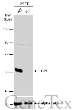
Wild-type (WT) and GPI knockout (KO) 293T cell extracts (30 μg) were separated by 7.5% SDS-PAGE, and the membrane was blotted with GPI antibody (GTX113203) diluted at 1:2000. The HRP-conjugated anti-rabbit IgG antibody (GTX213110-01) was used to detect the primary antibody, and the signal was developed with Trident ECL plus-Enhanced.
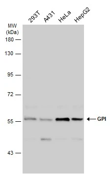
Various whole cell extracts (30 μg) were separated by 7.5% SDS-PAGE, and the membrane was blotted with GPI antibody (GTX113203) diluted at 1:2000. The HRP-conjugated anti-rabbit IgG antibody (GTX213110-01) was used to detect the primary antibody, and the signal was developed with Trident ECL plus-Enhanced.
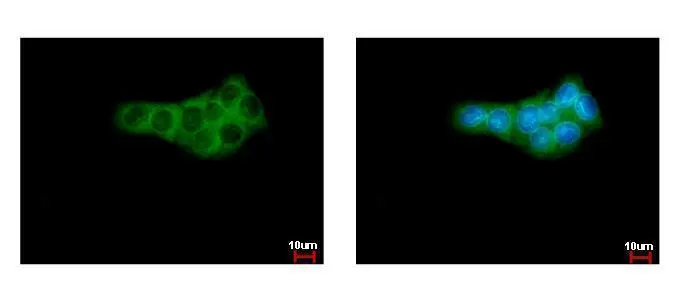
GPI antibody detects GPI protein at cytoplasm by immunofluorescent analysis.
Sample: HepG2 cells were fixed in ice-cold MeOH for 5 min.
Green: GPI protein stained by GPI antibody (GTX113203) diluted at 1:500.
Blue: Hoechst 33342 staining.
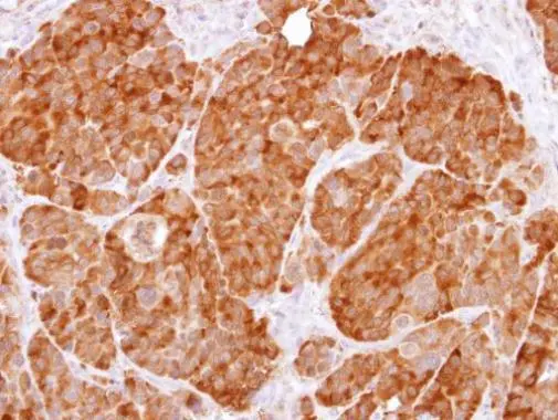
Immunohistochemical analysis of paraffin-embedded NCI-N87 xenograft, using GPI(GTX113203) antibody at 1:500 dilution.
Antigen Retrieval: Trilogy™ (EDTA based, pH 8.0) buffer, 15min
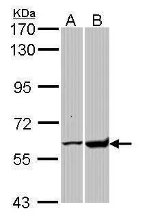
Sample (30 ug of whole cell lysate)
A: H1299
B: Raji
7.5% SDS PAGE
GTX113203 diluted at 1:10000
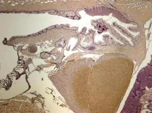
Immunohistochemical analysis of paraffin-embedded zebrafish tissue, using GPI antibody (GTX113203) at 1:300 dilution.
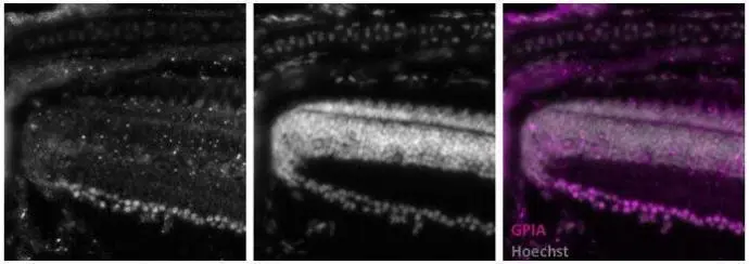
Immunohistochemical analysis of frozen sections of zebrafish retina, using GPI antibody (GTX113203) at 1:2000 dilution.
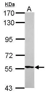
Sample (30 μg of whole cell lysate)
A: zebrafish eye
7.5% SDS PAGE
GTX113203 diluted at 1:1000
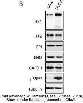
The data was published in the journal Viruses in 2018. PMID: 29518929
-
宿主Rabbit
-
克隆Polyclonal
-
同种型IgG
-
实验应用WB ICC/IF IHC-P IHC-Fr
-
种属反应Human, Zebrafish