HCN1抗体

HCN1 antibody detects HCN1 protein by immunohistochemical analysis.Sample: Paraffin-embedded mouse cerebellum.HCN1 stained by HCN1 antibody (GTX131334) diluted at 1:500.Antigen Retrieval: Citrate buffer, pH 6.0, 15 min

HCN1 antibody detects HCN1 protein by immunohistochemical analysis.Sample: Paraffin-embedded mouse hippocampus.HCN1 stained by HCN1 antibody (GTX131334) diluted at 1:500.Antigen Retrieval: Citrate buffer, pH 6.0, 15 min
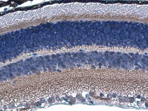
HCN1 antibody detects HCN1 protein in mouse retina by immunohistochemical analysis.
Sample: Paraffin-embedded mouse retina.
HCN1 antibody (GTX131334) diluted at 1:800.
Antigen Retrieval: Citrate buffer, pH 6.0, 15 min

HCN1 antibody detects HCN1 protein by immunofluorescent analysis.Sample: DIV9 rat E18 primary cortical neuron cells were fixed in 4% paraformaldehyde at RT for 15 min.Green: HCN1 stained by HCN1 antibody (GTX131334) diluted at 1:500.Red: beta Tubulin 3/ Tuj1, stained by beta Tubulin 3/ Tuj1 antibody [GT1338] (GTX631831) diluted at 1:500.Blue: Fluoroshield with DAPI (GTX30920).

HCN1 antibody detects HCN1 protein at cell membrane in rat brain by immunohistochemical analysis.
Sample: Paraffin-embedded rat brain.
HCN1 antibody (GTX131334) diluted at 1:500.
Antigen Retrieval: Citrate buffer, pH 6.0, 15 min
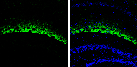
HCN1 antibody detects HCN1 protein expression by immunohistochemical analysis.
Sample: Frozen-sectioned adult mouse hippocampus.
Green: HCN1 protein stained by HCN1 antibody (GTX131334) diluted at 1:250.
Blue: Fluoroshield with DAPI (GTX30920).
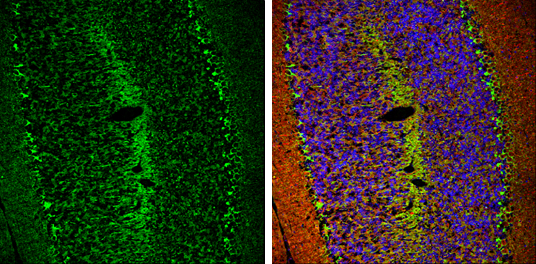
HCN1 antibody detects HCN1 protein expression by immunohistochemical analysis.
Sample: Frozen-sectioned adult mouse cerebellum.
Green: HCN1 protein stained by HCN1 antibody (GTX131334) diluted at 1:250.
Red: beta Tubulin 3/ TUJ1, stained by beta Tubulin 3/ TUJ1 antibody [GT11710] (GTX631836) diluted at 1:500.
Blue: Fluoroshield with DAPI (GTX30920).
-
宿主Rabbit
-
克隆Polyclonal
-
同种型IgG
-
实验应用ICC/IF IHC-P IHC-Fr
-
种属反应Mouse, Rat