HIF1 beta抗体

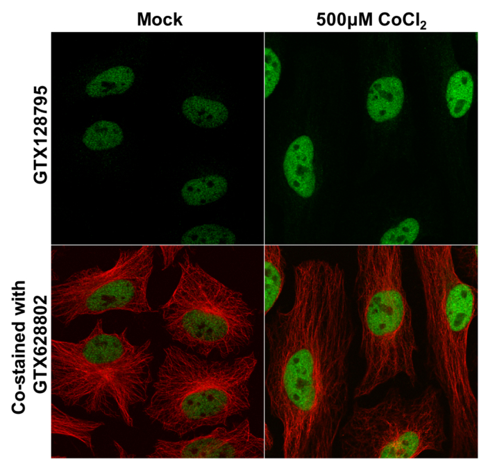
HIF1 beta antibody detects HIF1 beta protein at nucleus by immunofluorescent analysis.
Samples: HeLa cells mock (left) and treated with 500μM CoCl2 for 24hr (right) were fixed in 4% paraformaldehyde at RT for 15 min.
Green: HIF1 beta protein stained by HIF1 beta antibody (GTX128795) diluted at 1:500.
Red: alpha Tubulin, a cytoskeleton marker, stained by alpha Tubulin antibody [GT114] (GTX628802) diluted at 1:1000.
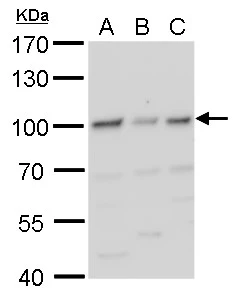
HIF1 beta antibody detects HIF1 beta protein by western blot analysis.
A. 30 μg 293T whole cell lysate/extract
B. 30 μg A431whole cell lysate/extract
C. 30 μg HeLa whole cell lysate/extract
7.5 % SDS-PAGE
HIF1 beta antibody (GTX128795) dilution: 1:1000
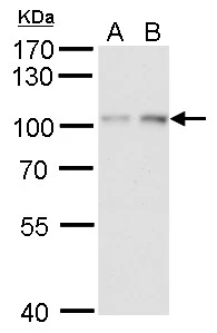
HIF1 beta antibody detects HIF1 beta protein by western blot analysis.
A. 30 μg HepG2 whole cell lysate/extract (untreated)
B. 30 μg HepG2 whole cell lysate/extract ( 1% O2 treatment for 24 hr)
7.5 % SDS-PAGE
HIF1 beta antibody (GTX128795) dilution: 1:1000
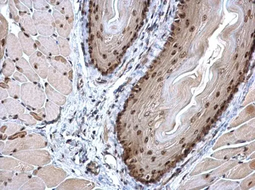
HIF1 beta antibody detects HIF1 beta protein at nucleus on mouse esophagus by immunohistochemical analysis.
Sample: Paraffin-embedded mouse esophagus.
HIF1 beta antibody (GTX128795) dilution: 1:500.
Antigen Retrieval: Trilogy™ (EDTA based, pH 8.0) buffer, 15min
-
宿主Rabbit
-
克隆Polyclonal
-
同种型IgG
-
实验应用WB ICC/IF IHC-P
-
种属反应Human, Mouse