Ku70抗体
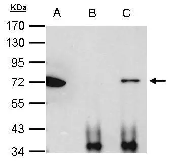
Ku70 antibody immunoprecipitates Ku70 protein in IP experiments. IP Sample: 1000 μg HeLa whole cell lysate/extract A. 40 μg HeLa whole cell lysate/extract B. Control with 2.5 μg of preimmune rabbit IgG C. Immunoprecipitation of Ku70 protein by 2.5 μg of Ku70 antibody (GTX101820) 7.5% SDS-PAGE The immunoprecipitated Ku70 protein was detected by Ku70 antibody (GTX101820) diluted at 1:1000. EasyBlot anti-rabbit IgG (GTX221666-01) was used as a secondary reagent.
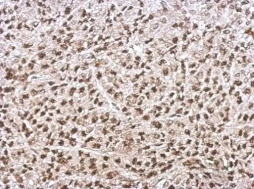
Immunohistochemical analysis of paraffin-embedded D54 xenograft, using Ku70(GTX101820) antibody at 1:500 dilution.
Antigen Retrieval: Trilogy™ (EDTA based, pH 8.0) buffer, 15min
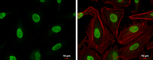
Ku70 antibody detects Ku70 protein at nucleus by immunofluorescent analysis.
Samples: HeLa cells were fixed in 4% paraformaldehyde at RT for 15 min.
Green: Ku70 protein stained by Ku70 antibody (GTX101820) diluted at 1:200.
Red: phalloidin, a cytoskeleton marker, diluted at 1:200.
Scale bar = 10 μm.
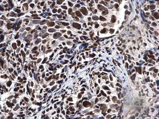
Ku70 antibody detects Ku70 protein at nucleus in human cervical cancer by immunohistochemical analysis.
Sample: Paraffin-embedded human cervical cancer.
Ku70 antibody (GTX101820) diluted at 1:500.
Antigen Retrieval: Citrate buffer, pH 6.0, 15 min
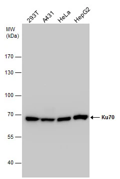
Ku70 antibody detects Ku70 protein by western blot analysis. Various whole cell extracts (30 μg) were separated by 7.5% SDS-PAGE, and the membrane was blotted with Ku70 antibody (GTX101820) diluted by 1:2000. The HRP-conjugated anti-rabbit IgG antibody (GTX213110-01) was used to detect the primary antibody.
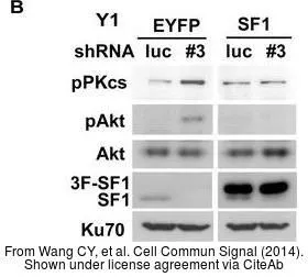
The data was published in the journal Cell Commun Signal in 2014. PMID: 25421435
-
宿主Rabbit
-
克隆Polyclonal
-
同种型IgG
-
实验应用WB ICC/IF IHC-P IP PLA
-
种属反应Human, Mouse