GeneTex中国官方网站
  • China

N-Cadherin抗体

Anti-N-Cadherin antibody used in Western Blot (WB). GTX127345
Anti-N-Cadherin antibody used in IHC (Frozen sections) (IHC-Fr). GTX127345
Anti-N-Cadherin antibody used in Western Blot (WB). GTX127345
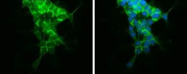
Anti-N-Cadherin antibody used in Immunocytochemistry/ Immunofluorescence (ICC/IF). GTX127345
Anti-N-Cadherin antibody used in Western Blot (WB). GTX127345
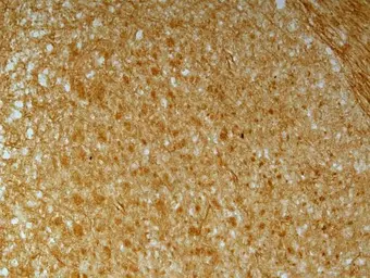
Anti-N-Cadherin antibody used in IHC (Frozen sections) (IHC-Fr). GTX127345
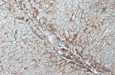
Anti-N-Cadherin antibody used in IHC (Paraffin sections) (IHC-P). GTX127345
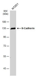
Anti-N-Cadherin antibody used in Western Blot (WB). GTX127345

目录号 GTX127345

宿主

Rabbit

克隆

Polyclonal

同种型

IgG

实验应用

WB, ICC/IF, IHC-P, IHC-Fr

种属反应

Human, Mouse, Rat, Monkey
产品包装
100 μl (¥4000),
25 μl (¥1700)
产品说明

摘要

N-Cadherin antibody recognizes N-cadherin protein, a calcium-dependent cell adhesion protein (predicted molecular weight of 100 kDa) encoded by the CDH2 gene. N-cadherin is involved in multiple processes, including neuron development and epithelial-mesenchymal transition (EMT). Due to the prominent role of EMT in cancer progression and metastasis, antibodies targeting either N-cadherin or E-cadherin are frequently used together to study the EMT process: the upregulation of N-cadherin followed by the downregulation of E-cadherin.

产品应用

应用说明

*最佳稀释倍数与浓度应由研究人员确认
实验应用 稀释倍数
WB 1:500-1:3000
ICC/IF 1:100-1:1000
IHC-P 1:100-1:1000
IHC-Fr 1:100-1:1000
WB: 免疫印迹
ICC/IF: 细胞染色
IHC-P: 石蜡切片
IHC-Fr: 冰冻切片

预期分子量

100 kDa. ( Note )

阳性对照

NT2D1 , SK-N-SH

预测种属反应

Bovine, Dog, Chicken, Xenopus tropicalis(>80% identity)

产品属性

形式

Liquid

存储溶液

PBS, 1% BSA, 20% Glycerol

存放说明

Store as concentrated solution. Centrifuge briefly prior to opening vial. For short-term storage (1-2 weeks), store at 4ºC. For long-term storage, aliquot and store at -20ºC or below. Avoid multiple freeze-thaw cycles.

浓度

0.15 mg/ml (Please refer to the vial label for the specific concentration.)

免疫原种属

Human

免疫原

Synthetic peptide encompassing a sequence within the Intracellular domain of human N-Cadherin. The exact sequence is proprietary.

纯化方式

Purified by antigen-affinity chromatography.

偶联

Unconjugated

RRID

AB_2885644

注意事项

仅供实验室使用。不适用于人类或动物的任何临床,治疗或诊断用途。不适合动物或人类食用。

相关讯息

别名

cadherin 2 , CD325 , CDHN , CDw325 , NCAD

细胞定位

Cell membrane

功能与背景

This gene is a classical cadherin from the cadherin superfamily. The encoded protein is a calcium dependent cell-cell adhesion glycoprotein comprised of five extracellular cadherin repeats, a transmembrane region and a highly conserved cytoplasmic tail. The protein functions during gastrulation and is required for establishment of left-right asymmetry. At certain central nervous system synapses, presynaptic to postsynaptic adhesion is mediated at least in part by this gene product. [provided by RefSeq]

数据库

研究领域

產品图片

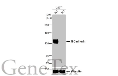
Anti-N-Cadherin antibody used in Western Blot (WB). GTX127345

GTX127345 WB Image

Wild-type (WT) and N-Cadherin knockout (KO) 293T cell extracts (30 μg) were separated by 5% SDS-PAGE, and the membrane was blotted with N-Cadherin antibody (GTX127345) diluted at 1:1000. The HRP-conjugated anti-rabbit IgG antibody (GTX213110-01) was used to detect the primary antibody.

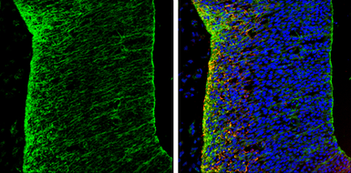
Anti-N-Cadherin antibody used in IHC (Frozen sections) (IHC-Fr). GTX127345

GTX127345 IHC-Fr Image

N-Cadherin antibody detects N-Cadherin protein expression by immunohistochemical analysis.
Sample: Frozen sectioned E13.5 Rat brain.
Green: N-Cadherin protein stained by N-Cadherin antibody (GTX127345) diluted at 1:250.
Red: beta Tubulin 3/ TUJ1, a mature neuron marker, stained by beta Tubulin 3/ TUJ1 antibody [GT11710] (GTX631836) diluted at 1:500.
Blue: Fluoroshield with DAPI (GTX30920).

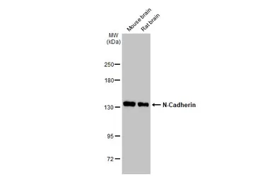
Anti-N-Cadherin antibody used in Western Blot (WB). GTX127345

GTX127345 WB Image

Various tissue extracts (50 μg) were separated by 5% SDS-PAGE, and the membrane was blotted with N-Cadherin antibody (GTX127345) diluted at 1:5000. The HRP-conjugated anti-rabbit IgG antibody (GTX213110-01) was used to detect the primary antibody.

Anti-N-Cadherin antibody used in Immunocytochemistry/ Immunofluorescence (ICC/IF). GTX127345

GTX127345 ICC/IF Image

N-Cadherin antibody detects N-Cadherin protein at cell membrane by immunofluorescent analysis.
Sample: SH-SY5Y cells were fixed in 4% paraformaldehyde at RT for 15 min.
Green: N-Cadherin protein stained by N-Cadherin antibody (GTX127345) diluted at 1:500.
Blue: Hoechst 33342 staining.

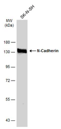
Anti-N-Cadherin antibody used in Western Blot (WB). GTX127345

GTX127345 WB Image

Whole cell extract (30 μg) was separated by 7.5% SDS-PAGE, and the membrane was blotted with N-Cadherin antibody (GTX127345) diluted at 1:1000. The HRP-conjugated anti-rabbit IgG antibody (GTX213110-01) was used to detect the primary antibody.

Anti-N-Cadherin antibody used in IHC (Frozen sections) (IHC-Fr). GTX127345

GTX127345 IHC-Fr Image

N-Cadherin antibody detects N-Cadherin protein on embryonic mouse brain by immunohistochemical analysis. Sample: Frozen section of embryonic mouse brain (mE18.5). N-Cadherin antibody (GTX127345) diluted at 1:500.

Anti-N-Cadherin antibody used in IHC (Paraffin sections) (IHC-P). GTX127345

GTX127345 IHC-P Image

N-Cadherin antibody detects N-Cadherin protein at cell membrane by immunohistochemical analysis.
Sample: Paraffin-embedded mouse liver.
N-Cadherin stained by N-Cadherin antibody (GTX127345) diluted at 1:500.
Antigen Retrieval: Citrate buffer, pH 6.0, 15 min

Anti-N-Cadherin antibody used in Western Blot (WB). GTX127345

GTX127345 WB Image

Whole cell extract (30 μg) was separated by 7.5% SDS-PAGE, and the membrane was blotted with N-Cadherin antibody (GTX127345) diluted at 1:1000. The HRP-conjugated anti-rabbit IgG antibody (GTX213110-01) was used to detect the primary antibody.

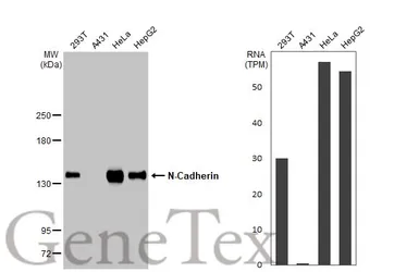
Anti-N-Cadherin antibody used in Western Blot (WB). GTX127345

GTX127345 WB Image

Various whole cell extracts (30 μg) were separated by 5% SDS-PAGE, and the membrane was blotted with N-Cadherin antibody (GTX127345) diluted at 1:1000. The HRP-conjugated anti-rabbit IgG antibody (GTX213110-01) was used to detect the primary antibody. Corresponding RNA expression data for the same cell lines are based on Human Protein Atlas program.

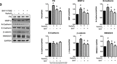
Anti-N-Cadherin antibody used in Western Blot (WB). GTX127345

GTX127345 WB Image

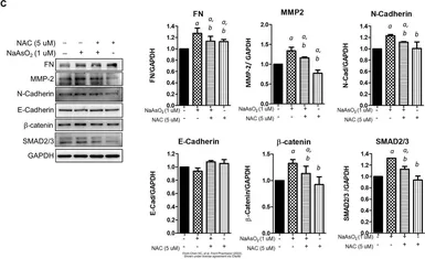
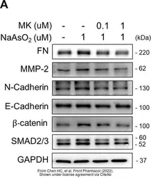
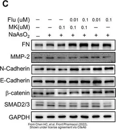
The data was published in the 2022 in Front Pharmacol. PMID: 35517780

Anti-N-Cadherin antibody used in Western Blot (WB). GTX127345

GTX127345 WB Image

The data was published in the journal EMBO Rep in 2020. PMID: 31908141

Anti-N-Cadherin antibody used in Western Blot (WB). GTX127345

GTX127345 WB Image

The data was published in the journal EMBO Rep in 2020. PMID: 31908141

Anti-N-Cadherin antibody used in Western Blot (WB). GTX127345

GTX127345 WB Image

The data was published in the journal EMBO Rep in 2020. PMID: 31908141

Anti-N-Cadherin antibody used in Western Blot (WB). GTX127345

GTX127345 WB Image

The data was published in the 2022 in Front Pharmacol. PMID: 35517780

Anti-N-Cadherin antibody used in Western Blot (WB). GTX127345

GTX127345 WB Image

The data was published in the 2022 in Front Pharmacol. PMID: 35517780

Anti-N-Cadherin antibody used in Western Blot (WB). GTX127345

GTX127345 WB Image

The data was published in the 2022 in Front Pharmacol. PMID: 35517780

文献引用

产品评论

N-Cadherin antibody 目录号 GTX127345

Rating ( Average 4.7 based on 3 users reviews)
实验应用
Western Blot(WB) ( Average 4.7 based on 3 users reviews)
Date : Anonymous submitted on 26-Nov-2019
Score :
Application Tested : WB
Sample Species : Rat
Sample : Membranes from cortical neurons
Amount used : 50 μg
Blocking : 5% milk, 24°C, 2Hr
Primary Antibody : 1:1000, 4°C, 16Hr
Date : Anonymous submitted on 29-Dec-2017
Score :
Application Tested : WB
Sample Species : Hu
Sample : FaDu xenograft
Amount used : 30 μg
Blocking : 4% non-fat milk in TBST, 23°C, 1Hr
Primary Antibody : 1:2000, 4°C, 16Hr
Date : Anonymous submitted on 05-Nov-2014
Score :
Application Tested : WB
Sample Species : Hu
Sample : A549 cells
Amount used : 20μg
Blocking : 5% skim milk in TBST, 24°C, 1Hr
Primary Antibody : 1:3000, 24°C, 15Hr
Application Reference
SDS
PBS.pdf
Glycerol.pdf
Proclin.pdf
产品包装 报价 (¥)
¥ 4000
¥ 1700
联系经销商
欣博盛生物科技有限公司
欣博盛生物科技有限公司
(全国热线) 4006-800-892 / (深圳) 0755-26755892 / (北京) 010-88594029 / (上海) 021-34613729 / (广州) 18024516375 / (香港) +852-69410778
http://www.neobioscience.com
genetex@neobioscience.com