Oct4抗体

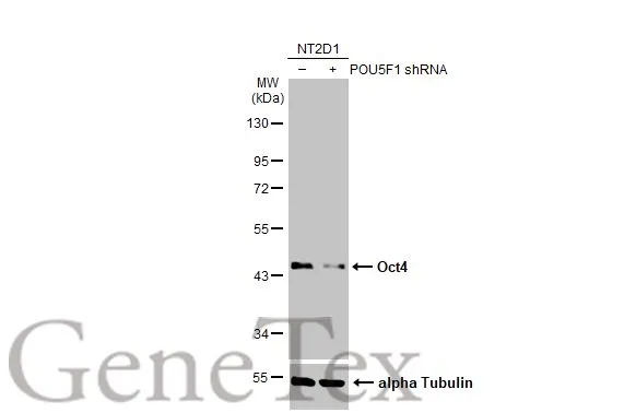
Non-transfected (–) and transfected (+) NT2D1 whole cell extracts (30 μg) were separated by 10% SDS-PAGE, and the membrane was blotted with Oct4 antibody (GTX100622) diluted at 1:10000. The HRP-conjugated anti-rabbit IgG antibody (GTX213110-01) was used to detect the primary antibody.
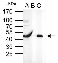
Oct4 antibody immunoprecipitates Oct4 protein in IP experiments. IP Sample: cell lysate/extract of Oct4 gene transfected 293T cells A. Cell lysate/extract of transfected 293T cell B. Control with 2 μg of preimmune rabbit IgG C. Immunoprecipitation of Oct4 by 2 μg of Oct4 antibody (GTX100622) 12% SDS-PAGE The immunoprecipitated Oct4 protein was detected by Oct4 antibody (GTX100622) diluted at 1:1000. EasyBlot anti-rabbit IgG (GTX221666-01) was used as a secondary reagent.
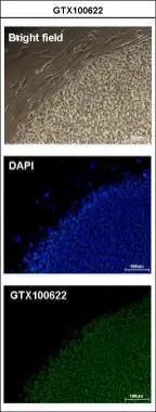
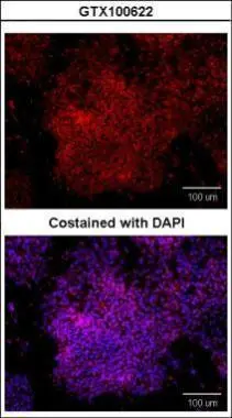
Immunofluorescence analysis of paraformaldehyde-fixed human embryonic stem cell, using Oct4(GTX100622) antibody at 1:200 dilution.
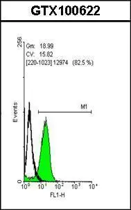
Flow cytometry on human embryonic stem cells, staining with Oct4 (GTX100622)antibody at 1:50 dilution(green) or rabbit IgG (black).
The observed M.W. is based on the publication: PMID: 23642364
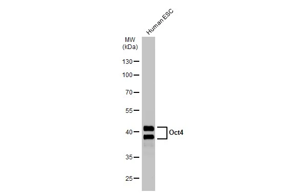
Human tissue extract (30 μg) was separated by 10% SDS-PAGE, and the membrane was blotted with Oct4 antibody (GTX100622) diluted at 1:5000. The HRP-conjugated anti-rabbit IgG antibody (GTX213110-01) was used to detect the primary antibody.
Sample (20 μg of whole cell lysate)
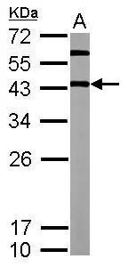
A: mouse ESC
12% SDS PAGE
GTX100622 diluted at 1:5000
The HRP-conjugated anti-rabbit IgG antibody (GTX213110-01) was used to detect the primary antibody.
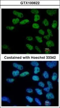
Immunofluorescence analysis of paraformaldehyde-fixed human embryonic stem cell, using Oct4(GTX100622) antibody at 1:100 dilution.
Immunofluorescence analysis of paraformaldehyde-fixed Human ESC, using Oct4(GTX100622) antibody at 1:400 dilution.
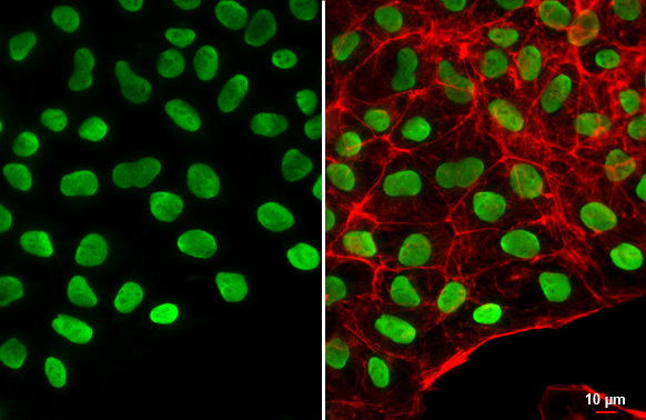
Oct4 antibody detects Oct4 protein at nucleus by immunofluorescent analysis.Sample: NT2D1 cells were fixed in 4% paraformaldehyde at RT for 15 min.Green: Oct4 stained by Oct4 antibody (GTX100622) diluted at 1:500.Red: phalloidin, a cytoskeleton marker, diluted at 1:200.Scale bar= 10 μm.
-
宿主Rabbit
-
克隆Polyclonal
-
同种型IgG
-
实验应用WB ICC/IF FCM IP
-
种属反应Human, Mouse