产品说明
摘要
PD-L1 antibody recognizes programmed death-ligand 1 (PD-L1) protein, also known as B7 homolog 1 (B7-H1) and cluster of differentiation 274 (CD274) protein. PD-L1 is found on various cancer cells and transduces immunosuppressive signals by binding to the programmed cell death protein 1 (PD-1) on effector T cells, thereby diminishing immune system attacks on malignant cells. Blocking the interaction between PD-L1 and PD-1 with PD-L1 antibodies is generating great clinical interest as a mode of immunotherapy against a number of cancers.
产品应用
应用说明
*最佳稀释倍数与浓度应由研究人员确认
实验应用 |
稀释倍数 |
1:500-1:3000 |
1:100-1:1000 |
1:100-1:1000 |
Assay dependent |
Assay dependent |
WB: 免疫印迹
ICC/IF: 细胞染色
IHC-P: 石蜡切片
IHC-Fr: 冰冻切片
预期分子量
Observed MW
50 kDa. The observed M.W. is based on the following publication. PMID: 23674495
阳性对照
MDA-MB-231 (24μg/ml Tunicamycin treatment for 16 hr) , MDA-MB-231 , A431(100 ng/mL IFN-gamma treatment for 48 hr)
产品属性
形式
Liquid
存储溶液
1XPBS, 1% BSA, 20% Glycerol (pH7)
存放说明
Store as concentrated solution. Centrifuge briefly prior to opening vial. For short-term storage (1-2 weeks), store at 4ºC. For long-term storage, aliquot and store at -20ºC or below. Avoid multiple freeze-thaw cycles.
浓度
0.29 mg/ml (Please refer to the vial label for the specific concentration.)
免疫原种属
Human
免疫原
Carrier-protein conjugated synthetic peptide encompassing a sequence within the C-terminus region of human PD-L1. The exact sequence is proprietary.
纯化方式
Purified by antigen-affinity chromatography.
偶联
Unconjugated
RRID
AB_1240586
注意事项
仅供实验室使用。不适用于人类或动物的任何临床,治疗或诊断用途。不适合动物或人类食用。
相关讯息
别名
CD274 molecule , B7-H , B7H1 , PD-L1 , PDCD1L1 , PDCD1LG1 , PDL1
细胞定位
Isoform 1: Cell membrane; Single-pass type I membrane protein , Isoform 2: Endomembrane system; Single-pass type I membrane protein
功能与背景
Involved in the costimulatory signal, essential for T-cell proliferation and production of IL10 and IFNG, in an IL2-dependent and a PDCD1-independent manner. Interaction with PDCD1 inhibits T-cell proliferation and cytokine production.
数据库
研究领域
產品图片
|
GTX104763 WB Image
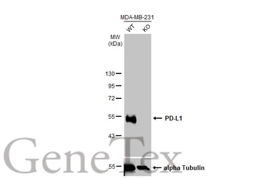
Wild-type (WT) and PD-L1 knockout (KO) MDA-MB-231 cell extracts (30 μg) were separated by 10% SDS-PAGE, and the membrane was blotted with PD-L1 antibody (GTX104763) diluted at 1:4000. The HRP-conjugated anti-rabbit IgG antibody (GTX213110-01) was used to detect the primary antibody.
|
|
GTX104763 WB Image
Untreated (–) and treated (+) A431 whole cell extracts (30 μg) were separated by 10% SDS-PAGE, and the membranes were blotted with PD-L1 antibody (GTX104763) diluted at 1:1200 and competitor's antibody (CST#13684) diluted at 1:500. The HRP-conjugated anti-rabbit IgG antibody (GTX213110-01) was used to detect the primary antibody.
*The competitor is not affiliated with GeneTex and does not endorse this product.
|
|
GTX104763 ICC/IF Image
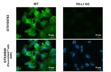
PD-L1 antibody detects PD-L1 protein at cell membrane by immunofluorescent analysis.Sample: MDA-MB-231 cells were fixed in ice-cold MeOH for 5 min.Green: PD-L1 stained by PD-L1 antibody (GTX104763) diluted at 1:500.Blue: Fluoroshield with DAPI (GTX30920).
|
|
GTX104763 WB Image
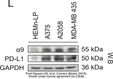
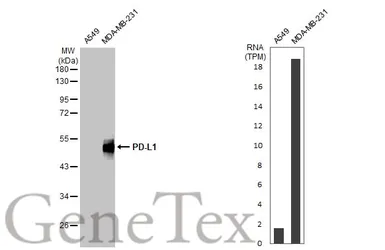
Various whole cell extracts (30 μg) were separated by 10% SDS-PAGE, and the membrane was blotted with PD-L1 antibody (GTX104763) diluted at 1:2000. The HRP-conjugated anti-rabbit IgG antibody (GTX213110-01) was used to detect the primary antibody, and the signal was developed with Trident ECL plus-Enhanced.
|
|
GTX104763 WB Image
Various whole cell extracts (30 μg) were separated by 10% SDS-PAGE, and the membrane was blotted with PD-L1 antibody (GTX104763) diluted at 1:2000. The HRP-conjugated anti-rabbit IgG antibody (GTX213110-01) was used to detect the primary antibody.
|
|
GTX104763 WB Image
Non-transfected (–) and transfected (+) A431 whole cell extracts (30 μg) were separated by 10% SDS-PAGE, and the membrane was blotted with PD-L1 antibody (GTX104763) diluted at 1:1000. The HRP-conjugated anti-rabbit IgG antibody (GTX213110-01) was used to detect the primary antibody.
|
|
GTX104763 WB Image
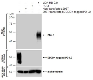
Various whole cell extracts were separated by 10% SDS-PAGE, and the membranes were blotted with PD-L1 antibody (GTX104763) diluted at 1:600 and with DDDDK tag antibody (GTX115043) diluted at 1:3000 to detect DDDDK-tagged PD-L2. The HRP-conjugated anti-rabbit IgG antibody (GTX213110-01) was used to detect the primary antibody.
|
|
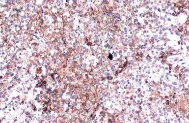
GTX104763 IHC-P Image
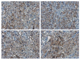
PD-L1 antibody detects PD-L1 protein at cell membrane by immunohistochemical analysis.Sample: Paraffin-embedded human ovarian cancer.PD-L1 stained by PD-L1 antibody (GTX104763) diluted at 1:4000.Antigen Retrieval: Citrate buffer, pH 6.0, 15 min
|
|
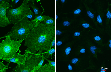
GTX104763 ICC/IF Image
PD-L1 antibody detects PD-L1 protein by immunofluorescent analysis.Sample: MDA-MB-231 (left) and HeLa (right) cells were fixed in ice-cold MeOH for 5 min.Green: PD-L1 stained by PD-L1 antibody (GTX104763) diluted at 1:500.Blue: Hoechst 33342 staining.Scale bar= 10 μm.
|
|
GTX104763 IHC-P Image
PD-L1 antibody detects PD-L1 proteinat cell membrane in human ovarian carcinoma by immunohistochemical analysis. Sample: Paraffin-embedded human ovarian carcinoma. PD-L1 antibody (GTX104763) diluted at 1:1000.
Antigen Retrieval: Citrate buffer, pH 6.0, 15 min
|
|
GTX104763 WB Image
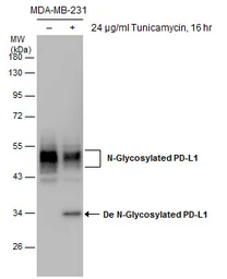
Untreated (–) and treated (+) MDA-MB-231 whole cell extracts (30 μg) were separated by 10% SDS-PAGE, and the membrane was blotted with PD-L1 antibody (GTX104763) diluted at 1:1000.
|
|
GTX104763 WB Image
Various whole cell extracts (30 μg) were separated by 12% SDS-PAGE, and the membranes were blotted with PD-L1 antibody (GTX104763) diluted at 1:2000 and competitor's antibody (CST#13684) diluted by 1:500. The HRP-conjugated anti-rabbit IgG antibody (GTX213110-01) was used to detect the primary antibody.
*The competitor is not affiliated with GeneTex and does not endorse this product.
|
|
GTX104763 IHC-P Image
PD-L1 antibody detects PD-L1 protein at cell membrane in PD-L1 protein-expressing cell lines by immunohistochemical analysis. Antibodies: PD-L1 antibody (GTX104763) diluted at 1:1000, and competitor's antibody diluted at 1:50. Samples: Negative (-), low positive (+), intermediate positive (++) and strong positive (+++) cell line cores assessed using Quantitative Digital Pathology.
Antigen Retrieval: Citrate buffer, pH 6.0, 15 min
*The competitor is not affiliated with GeneTex and does not endorse this product.
|
|
GTX104763 IHC-P Image
PD-L1 antibody detects PD-L1 protein at cell membrane in human ovarian carcinoma by immunohistochemical analysis. Antibodies: PD-L1 antibody (GTX104763) diluted at 1:1000, and competitor's antibody diluted at 1:50.
Antigen Retrieval: Citrate buffer, pH 6.0, 15 min
*The competitor is not affiliated with GeneTex and does not endorse this product.
|
|
GTX104763 WB Image
Non-transfected (–) and transfected (+) 293T whole cell extracts (30 μg) were separated by 10% SDS-PAGE, and the membrane was blotted with PD-L1 antibody (GTX104763) diluted at 1:1000. The HRP-conjugated anti-rabbit IgG antibody (GTX213110-01) was used to detect the primary antibody, and the signal was developed with Trident ECL plus-Enhanced.
|
文献引用
产品评论
PD-L1 antibody
目录号 GTX104763
Rating
|
|
( Average 4.8 based on 11 users reviews)
|
实验应用
Western Blot(WB)
|
|
( Average 4.8 based on 11 users reviews)
|
Date :
Anonymous submitted on 24-Jul-2023
Application Tested :
WB
Sample Species :
Hu
Figure Note :
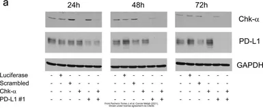
The data was peer reviewed and published in the 2021 in Cancer Metab.
PMID: 33608051
Date :
Anonymous submitted on 10-Aug-2021
Application Tested :
WB
Sample Species :
Hu
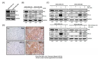
Figure Note :
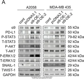
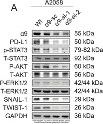
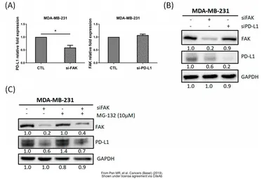
The data was peer reviewed and published in the journal Cancers (Basel) in 2019.
PMID: 31835799
Date :
Anonymous submitted on 10-Aug-2021
Application Tested :
WB
Sample Species :
Hu
Figure Note :
The data was peer reviewed and published in the journal Cancers (Basel) in 2019.
PMID: 31835799
Date :
Anonymous submitted on 10-Aug-2021
Application Tested :
WB
Sample Species :
Hu
Figure Note :
The data was peer reviewed and published in the journal Cancers (Basel) in 2019.
PMID: 31835799
Date :
Anonymous submitted on 10-Aug-2021
Application Tested :
WB
Sample Species :
Hu
Figure Note :
The data was peer reviewed and published in the journal Cancers (Basel) in 2019.
PMID: 31835799
Date :
Anonymous submitted on 10-Aug-2021
Application Tested :
WB
Sample Species :
Hu
Figure Note :
The data was peer reviewed and published in the journal Cancers (Basel) in 2019.
PMID: 31905966
Date :
Anonymous submitted on 10-Aug-2021
Application Tested :
WB
Sample Species :
Hu
Figure Note :
The data was peer reviewed and published in the journal Cancers (Basel) in 2019.
PMID: 31905966
Date :
Anonymous submitted on 09-May-2021
Application Tested :
WB
Sample Species :
Hu
Sample :
Huh7 cells
Amount used :
10 μg
Blocking :
5% skim milk in TBST, 27°C, 1Hr
Primary Antibody :
1:5000, 4°C, 12Hr
Secondary Antibody :
GTX213110-01, 1:5000, 27°C, 2Hr
条带说明 :
Two groups (regorafenib -/+) of whole cell extracts (10 μg) were separated by 12% SDS-PAGE, and the membrane was blotted with PD-L1 antibody (GTX104763) diluted at 1:5000. The HRP-conjugated anti-rabbit IgG antibody (GTX213110-01) was used to detect the primary antibody.
Date :
Anonymous submitted on 18-Mar-2021
Application Tested :
WB
Sample Species :
Hu
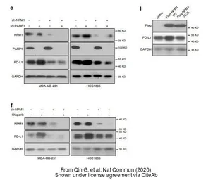
Figure Note :
The data was peer reviewed and published in the journal Nat Commun in 2020.
PMID: 32245950
Date :
Anonymous submitted on 18-Mar-2021
Application Tested :
WB
Sample Species :
Hu
Figure Note :
The data was peer reviewed and published in the journal Nat Commun in 2020.
PMID: 32245950
Date :
Anonymous submitted on 22-May-2017
Application Tested :
WB
Sample Species :
Hu
Sample :
SK-LMS-1 cells
Amount used :
30 μg
Blocking :
3%BSA, 25°C, 0.5Hr
Primary Antibody :
1:1000, 4°C, 16Hr