PD1抗体
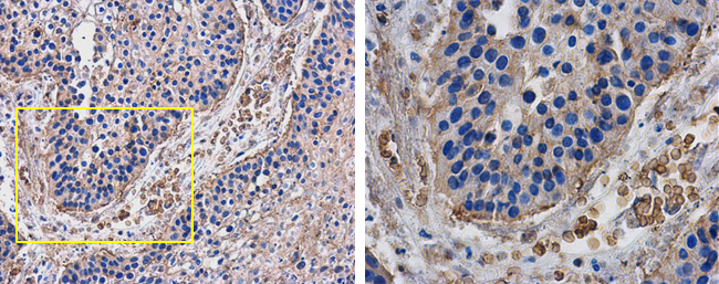
PD1 antibody detects PD1 protein at cell membrane of inflammatory cells in human lung cancer by immunohistochemical analysis.
Sample: Paraffin-embedded human lung cancer.
PD1 antibody (GTX128435) diluted at 1:500.
Antigen Retrieval: Citrate buffer, pH 6.0, 15 min
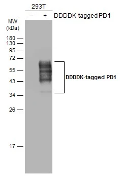
Non-transfected (–) and transfected (+) 293T whole cell extracts (30 μg) were separated by 12% SDS-PAGE, and the membrane was blotted with PD1 antibody (GTX128435) diluted at 1:5000. The HRP-conjugated anti-rabbit IgG antibody (GTX213110-01) was used to detect the primary antibody.
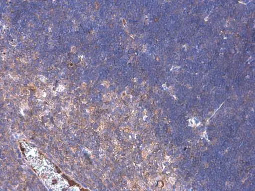
PD1 antibody detects PD1 protein at cell membrane by immunohistochemical analysis.Sample: Paraffin-embedded mouse thymus gland.PD1 stained by PD1 antibody (GTX128435) diluted at 1:500.
Antigen Retrieval: Citrate buffer, pH 6.0, 15 min
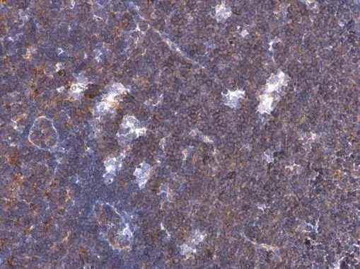
PD1 antibody detects PD1 protein at cell membrane by immunohistochemical analysis.Sample: Paraffin-embedded mouse lymph node.PD1 stained by PD1 antibody (GTX128435) diluted at 1:500.
Antigen Retrieval: Citrate buffer, pH 6.0, 15 min
-
宿主Rabbit
-
克隆Polyclonal
-
同种型IgG
-
实验应用WB IHC-P
-
种属反应Human, Mouse