RPS6抗体
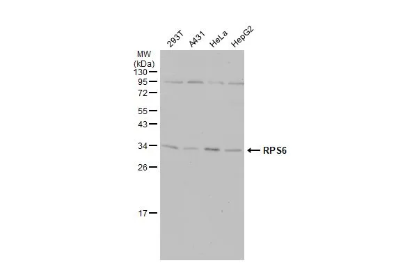
Various whole cell extracts (30 μg) were separated by 12% SDS-PAGE, and the membrane was blotted with RPS6 antibody (GTX113542) diluted at 1:500. The HRP-conjugated anti-rabbit IgG antibody (GTX213110-01) was used to detect the primary antibody.
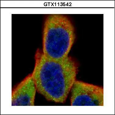
Confocal immunofluorescence analysis (Olympus FV10i) of paraformaldehyde-fixed A431, using RPS6(GTX113542) antibody (Green) at 1:500 dilution. Alpha-tubulin filaments were labeled with GTX11304 (Red) at 1:500.
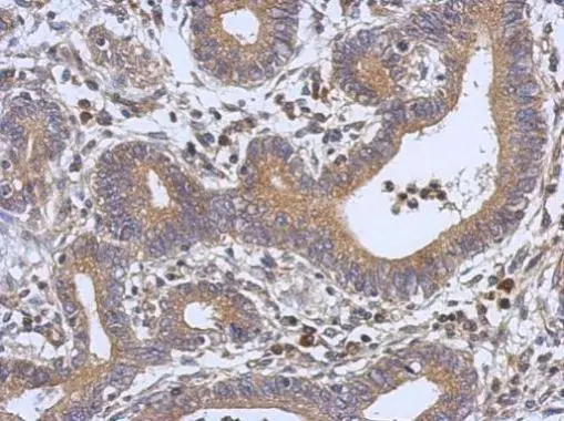
Immunohistochemical analysis of paraffin-embedded human colon carcinoma, using RPS6(GTX113542) antibody at 1:500 dilution.
Antigen Retrieval: Trilogy™ (EDTA based, pH 8.0) buffer, 15min
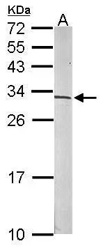
Sample (50 μg of whole cell lysate)
A: mouse brain
12% SDS PAGE
GTX113542 diluted at 1:500
The HRP-conjugated anti-rabbit IgG antibody (GTX213110-01) was used to detect the primary antibody.
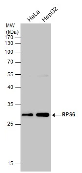
Various whole cell extracts (30 μg) were separated by 12% SDS-PAGE, and the membrane was blotted with RPS6 antibody (GTX113542) diluted at 1:500. The HRP-conjugated anti-rabbit IgG antibody (GTX213110-01) was used to detect the primary antibody.
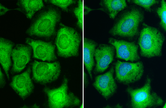
RPS6 antibody detects RPS6 protein at cytoplasm and nucleus by immunofluorescent analysis.Sample: HeLa cells were fixed in 4% paraformaldehyde at RT for 15 min.Green: RPS6 stained by RPS6 antibody (GTX113542) diluted at 1:500.Blue: Hoechst 33342 staining.
-
宿主Rabbit
-
克隆Polyclonal
-
同种型IgG
-
实验应用WB ICC/IF IHC-P IHC-Wm
-
种属反应Human, Mouse, Zebrafish