SP1抗体


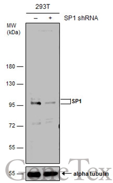
Non-transfected (–) and transfected (+) 293T whole cell extracts (50 μg) were separated by 7.5% SDS-PAGE, and the membrane was blotted with SP1 antibody (GTX110593) diluted at 1:10000. The HRP-conjugated anti-rabbit IgG antibody (GTX213110-01) was used to detect the primary antibody, and the signal was developed with Trident ECL plus-Enhanced.
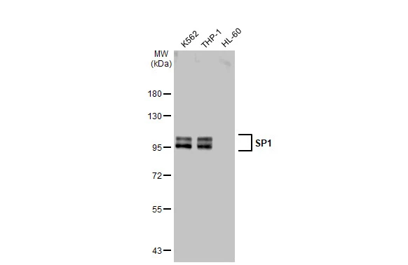
Various whole cell extracts (30 μg) were separated by 7.5% SDS-PAGE, and the membrane was blotted with SP1 antibody (GTX110593) diluted at 1:2000. The HRP-conjugated anti-rabbit IgG antibody (GTX213110-01) was used to detect the primary antibody.
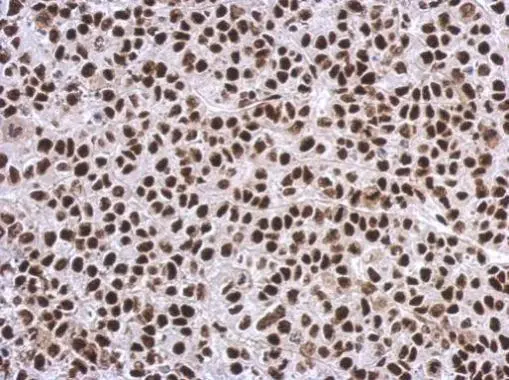
Immunohistochemical analysis of paraffin-embedded Hela xenograft, using SP1(GTX110593) antibody at 1:500 dilution.
Antigen Retrieval: Trilogy™ (EDTA based, pH 8.0) buffer, 15min
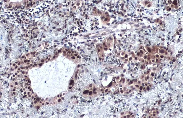
SP1 antibody detects SP1 protein at cytoplasm and nucleus by immunohistochemical analysis.Sample: Paraffin-embedded human lung cancer.SP1 stained by SP1 antibody (GTX110593) diluted at 1:2000.Antigen Retrieval: Citrate buffer, pH 6.0, 15 min
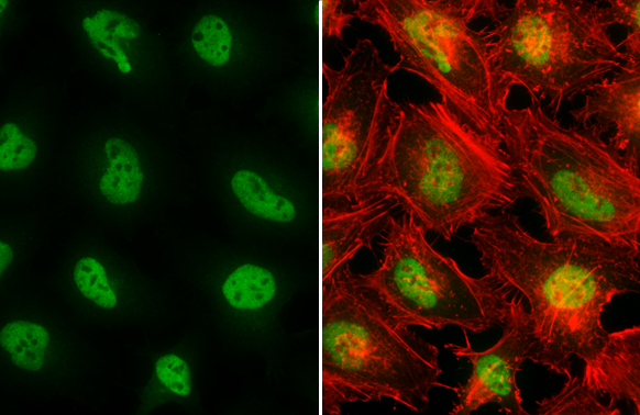
SP1 antibody detects SP1 protein at nucleus by immunofluorescent analysis.Sample: HeLa cells were fixed in 4% paraformaldehyde at RT for 15 min.Green: SP1 stained by SP1 antibody (GTX110593) diluted at 1:1000.Red: phalloidin, a cytoskeleton marker, diluted at 1:200.
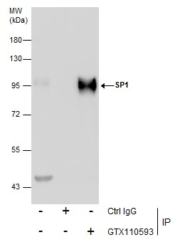
Immunoprecipitation of SP1 protein from THP-1 whole cell extracts using 5 μg of OCT1 antibody (GTX110593).
Western blot analysis was performed using OCT1 antibody (GTX110593) diluted at 1:250.
EasyBlot anti-Rabbit IgG (GTX221666-01) was used as a secondary reagent.
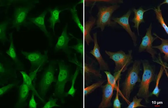
SP1 antibody detects SP1 protein at nucleus by immunofluorescent analysis.Sample: HeLa cells were fixed in 4% paraformaldehyde at RT for 15 min.Green: SP1 stained by SP1 antibody (GTX110593) diluted at 1:100.Red: alpha Tubulin, a cytoskeleton marker, stained by alpha Tubulin antibody [GT114] (GTX628802) diluted at 1:1000.Blue: Fluoroshield with DAPI (GTX30920).
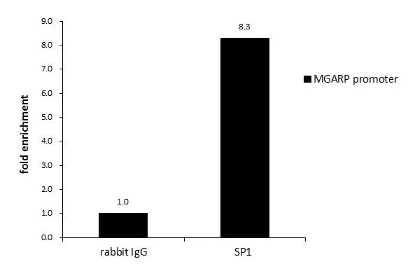
ChIP was performed with 293T chromatin extract and 5 μg of either normal rabbit IgG or anti-SP1 antibody. The precipitated DNA was detected by PCR with primer set targeting to MGARP promoter.
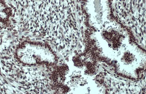
SP1 antibody detects SP1 protein at nucleus by immunohistochemical analysis.Sample: Paraffin-embedded human breast carcinoma.SP1 stained by SP1 antibody (GTX110593) diluted at 1:500.Antigen Retrieval: Citrate buffer, pH 6.0, 15 min
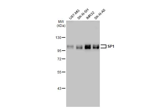
Various whole cell extracts (30 μg) were separated by 7.5% SDS-PAGE, and the membrane was blotted with SP1 antibody (GTX110593) diluted at 1:2000. The HRP-conjugated anti-rabbit IgG antibody (GTX213110-01) was used to detect the primary antibody.
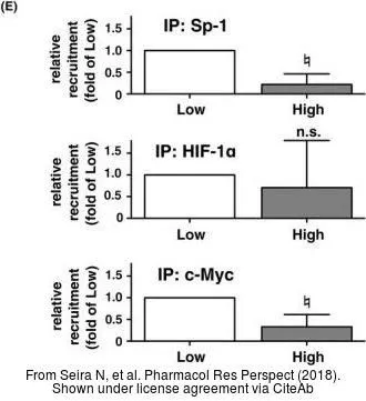
The data was published in the journal Pharmacol Res Perspect in 2018. PMID: 30455960
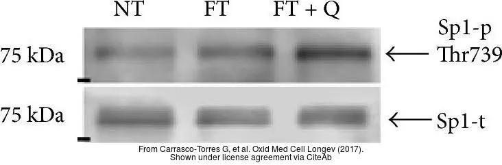
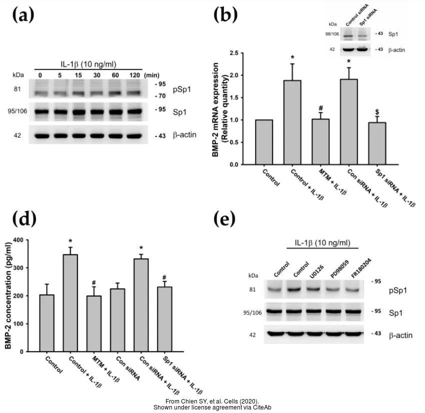
The data was published in the 2017 in Oxid Med Cell Longev. PMID: 28740570
-
宿主Rabbit
-
克隆Polyclonal
-
同种型IgG
-
实验应用WB ICC/IF IHC-P IP ChIP assay
-
种属反应Human