Synaptophysin抗体


Immunohistochemical characterization of Synaptophysin (GTX100865), p63 (GTX102425) and Cytokeratin 7 (GTX109723) in human small cell lung cancer (SCLC) and non-small cell lung cancer (NSCLC) specimens.
Sample: Paraffin-embedded human SCLC (upper panel) and NSCLC (lower panel).
The section was pre-treated using heat mediated antigen retrieval with sodium citrate buffer (pH6) for 15 mins. The section was then incubated with primary antibody at 1:500 overnight at 4ºC and detected using an HRP conjugated avidin-biotin-peroxidase Complex system. DAB was used as the chromogen and counterstained with haematoxylin.
Antigen Retrieval: Citrate buffer, pH 6.0, 15 min
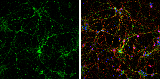
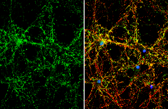
Synaptophysin antibody detects Synaptophysin protein at synaptic vesicles by immunofluorescent analysis.
Sample: DIV9 rat E18 primary cortical neurons were fixed in 4% paraformaldehyde at RT for 15 min.
Green: Synaptophysin protein stained by Synaptophysin antibody (GTX100865) diluted at 1:500.
Red: beta Tubulin 3/ Tuj1, stained by beta Tubulin 3/ Tuj1 antibody [GT11710] (GTX631836) diluted at 1:500.
Blue: Fluoroshield with DAPI (GTX30920).
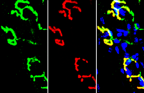
Synaptophysin antibody detects Synaptophysin protein by immunohistochemical analysis.Sample: Frozen-sectioned mouse muscle.Green: Synaptophysin stained by Synaptophysin antibody (GTX100865) diluted at 1:250.Red: α-Bungarotoxin, stained by α-Bungarotoxin, Alexa Fluor™ 594 conjugate (B13423) diluted at 1:5000.Blue: Hoechst 33342 staining.
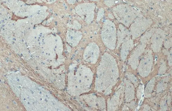
Synaptophysin antibody detects Synaptophysin protein at cell membrane and cytoplasm by immunohistochemical analysis.Sample: Paraffin-embedded mouse brain.Synaptophysin stained by Synaptophysin antibody (GTX100865) diluted at 1:500.Antigen Retrieval: Citrate buffer, pH 6.0, 15 min
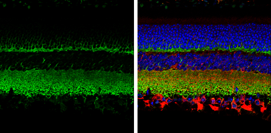
Synaptophysin antibody detects Synaptophysin protein expression by immunohistochemical analysis.
Sample: Paraffin-Embedded adult mouse retina.
Green: Synaptophysin protein stained by Synaptophysin antibody (GTX100865) diluted at 1:250.
Red: beta Tubulin 3/ TUJ1, stained by beta Tubulin 3/ TUJ1 antibody [GT11710] (GTX631836) diluted at 1:250.
Blue: Fluoroshield with DAPI (GTX30920).
Antigen Retrieval: Citrate buffer, pH 6.0, 15 min
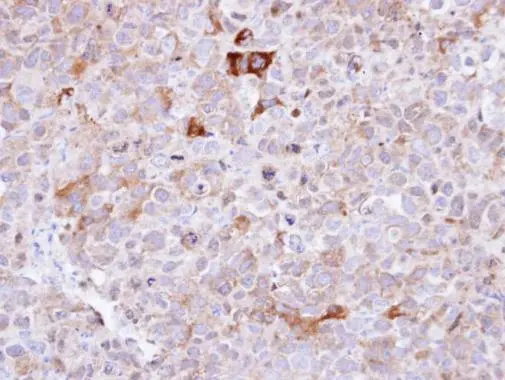
Immunohistochemical analysis of paraffin-embedded CL1-0 xenograft , using Synaptophysin(GTX100865) antibody at 1:100 dilution.
Antigen Retrieval: Trilogy™ (EDTA based, pH 8.0) buffer, 15min
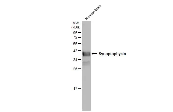
Human tissue extract (30 μg) was separated by 12% SDS-PAGE, and the membrane was blotted with Synaptophysin antibody (GTX100865) diluted at 1:50000. The HRP-conjugated anti-rabbit IgG antibody (GTX213110-01) was used to detect the primary antibody.
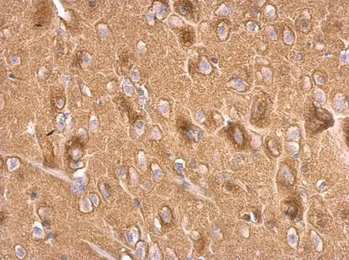
Synaptophysin antibody detects Synaptophysin protein at cell membrane and cytoplasm by immunohistochemical analysis.Sample: Paraffin-embedded rat brain.Synaptophysin stained by Synaptophysin antibody (GTX100865) diluted at 1:500.Antigen Retrieval: Citrate buffer, pH 6.0, 15 min
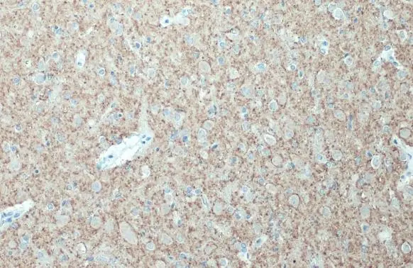
Synaptophysin antibody detects Synaptophysin protein at on rat fore brain by immunohistochemical analysis.
Sample: Paraffin-embedded rat fore brain.
Synaptophysin antibody (GTX100865) dilution: 1:500.
Antigen Retrieval: Trilogy™ (EDTA based, pH 8.0) buffer, 15min
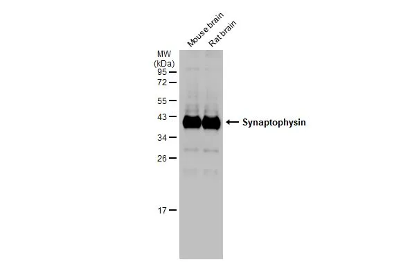
Various tissue extracts (50 μg) were separated by 12% SDS-PAGE, and the membrane was blotted with Synaptophysin antibody (GTX100865) diluted at 1:50000. The HRP-conjugated anti-rabbit IgG antibody (GTX213110-01) was used to detect the primary antibody.
Synaptophysin antibody detects Synaptophysin protein by immunofluorescent analysis.Sample: DIV10 rat E18 primary hippocampal neuron cells were fixed in 4% paraformaldehyde at RT for 15 min.Green: Synaptophysin stained by Synaptophysin antibody (GTX100865) diluted at 1:500.Red: Tau, stained by Tau antibody [GT287] (GTX634809) diluted at 1:500.Blue: Fluoroshield with DAPI (GTX30920).
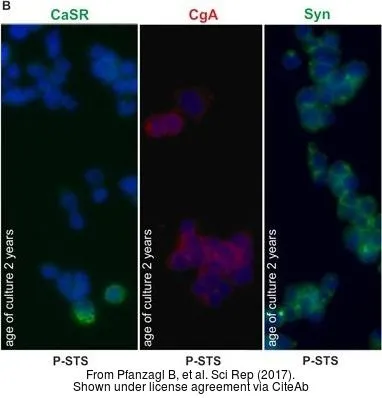
The data was published in the journal Sci Rep in 2017.PMID: 28465562
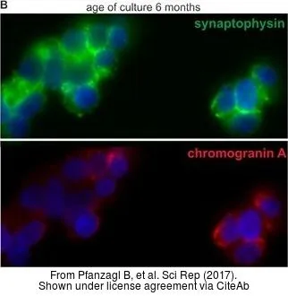
The data was published in the journal Sci Rep in 2017.PMID: 28465562
-
宿主Rabbit
-
克隆Polyclonal
-
同种型IgG
-
实验应用WB ICC/IF IHC-P IHC-Fr IHC-Wm
-
种属反应Human, Mouse, Rat