TNF alpha抗体


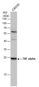
TNF alpha antibody detects TNF alpha protein by western blot analysis. Whole cell extracts (30 μg) was separated by 12% SDS-PAGE, and the membrane was blotted with TNF alpha antibody (GTX110520) diluted at 1:500. The HRP-conjugated anti-rabbit IgG antibody (GTX213110-01) was used to detect the primary antibody.
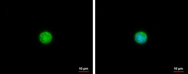
TNF alpha antibody detects TNF alpha protein at cytoplasm by immunofluorescent analysis.
Sample: Raw264.7 cells were fixed in 4% paraformaldehyde at RT for 15 min.
Green: TNF alpha protein stained by TNF alpha antibody (GTX110520) diluted at 1:500.
Blue: Hoechst 33342 staining.
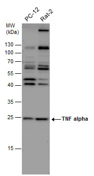
TNF alpha antibody detects TNF alpha protein by western blot analysis. Various whole cell extracts (30 μg) were separated by 12% SDS-PAGE, and the membrane was blotted with TNF alpha antibody (GTX110520) diluted at 1:500. The HRP-conjugated anti-rabbit IgG antibody (GTX213110-01) was used to detect the primary antibody.
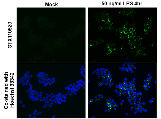
TNF alpha antibody detects TNF alpha protein at membrane by immunofluorescent analysis.
Sample: jurkat cells were fixed in 4% paraformaldehyde at RT for 15 min.
Green: TNF alpha protein stained by TNF alpha antibody (GTX110520) diluted at 1:500.
Blue: Hoechst 33342 staining.
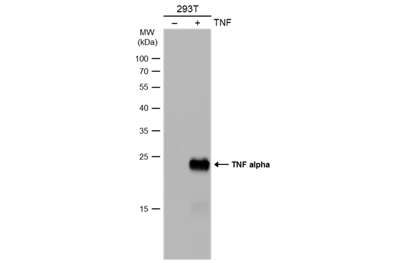
Non-transfected (–) and transfected (+) 293T whole cell extracts (30 μg) were separated by 12% SDS-PAGE, and the membrane was blotted with TNF alpha antibody (GTX110520) diluted at 1:1000. The HRP-conjugated anti-rabbit IgG antibody (GTX213110-01) was used to detect the primary antibody.
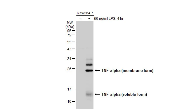
Untreated (–) and treated (+) Raw264.7 whole cell extracts (30 μg) were separated by 12% SDS-PAGE, and the membrane was blotted with TNF alpha antibody (GTX110520) diluted at 1:1000. The HRP-conjugated anti-rabbit IgG antibody (GTX213110-01) was used to detect the primary antibody.
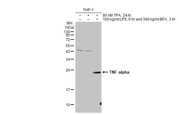
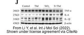
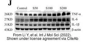
Untreated (–) and treated (+) THP-1 whole cell extracts (30 μg) were separated by 12% SDS-PAGE, and the membrane was blotted with TNF alpha antibody (GTX110520) diluted at 1:500. The HRP-conjugated anti-rabbit IgG antibody (GTX213110-01) was used to detect the primary antibody.
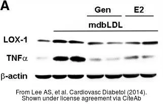
The data was published in the journal Cardiovasc Diabetol in 2014. PMID: 24666525
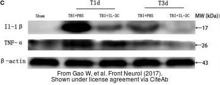
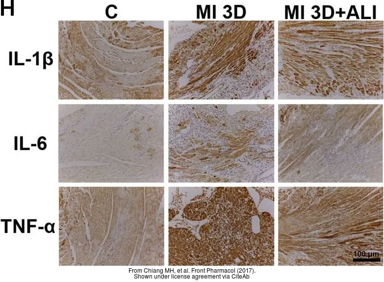
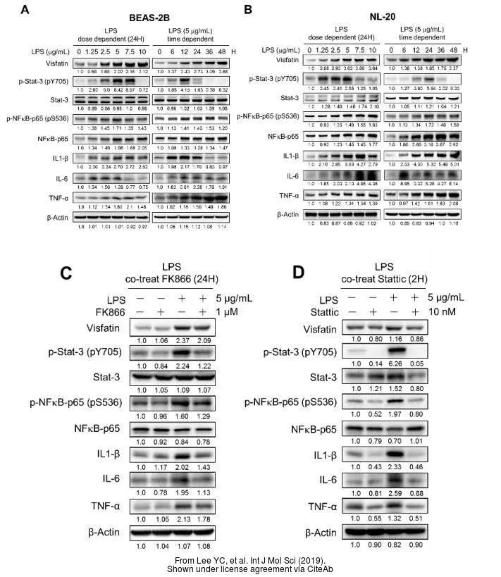
The data was published in the journal Front Pharmacol in 2017.PMID: 29184499
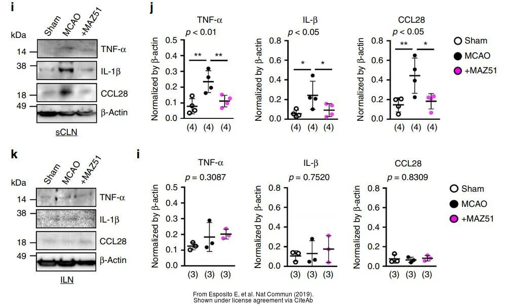
The data was published in the journal Nat Commun in 2019.PMID: 31757960
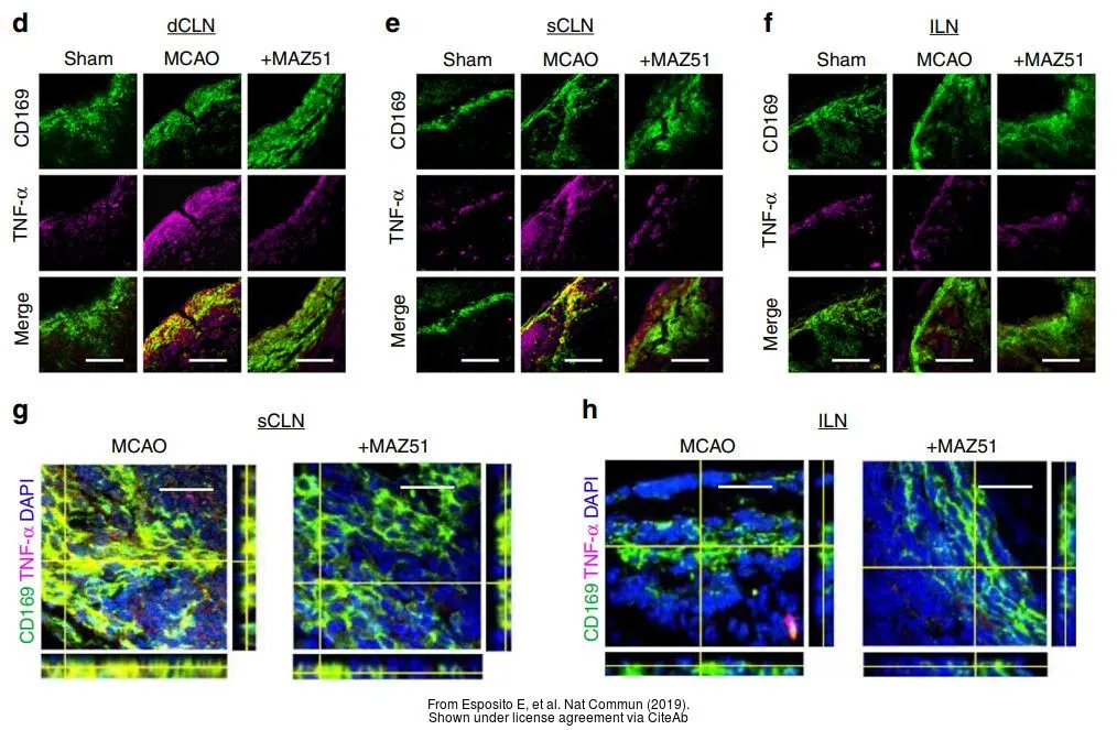
The data was published in the journal Nat Commun in 2019.PMID: 31757960
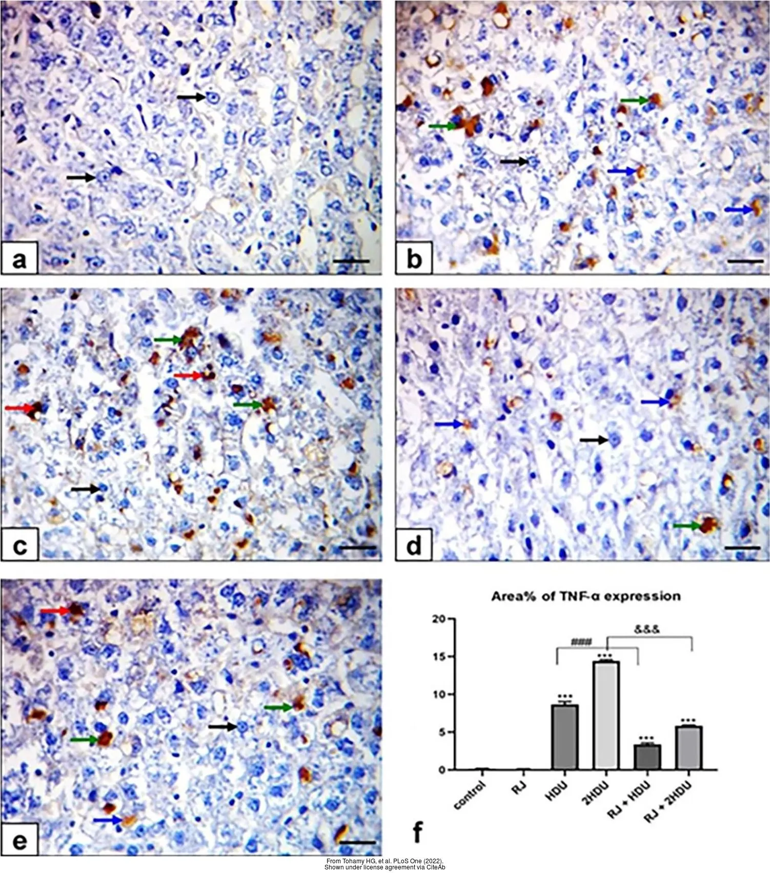
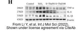
The data was published in the 2022 in PLoS One. PMID: 35303036
-
宿主Rabbit
-
克隆Polyclonal
-
同种型IgG
-
实验应用WB ICC/IF IHC-P IHC-Fr
-
种属反应Human, Mouse, Rat, Bovine