Tau抗体


Non-transfected (–) and transfected (+) SK-N-SH whole cell extracts (30 μg) were separated by 10% SDS-PAGE, and the membrane was blotted with Tau antibody (GTX130462) diluted at 1:500. The HRP-conjugated anti-rabbit IgG antibody (GTX213110-01) was used to detect the primary antibody, and the signal was developed with Trident ECL plus-Enhanced.
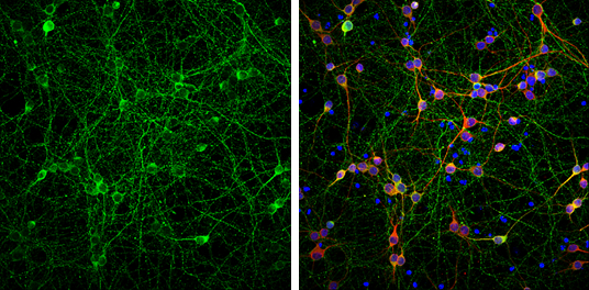
Tau antibody detects Tau protein at axon by immunofluorescent analysis.
Sample: DIV9 rat E18 primary cortical neurons were fixed in 4% paraformaldehyde at RT for 15 min.
Green: Tau protein stained by Tau antibody (GTX130462) diluted at 1:500.
Red: MAP2, stained by MAP2 antibody [HM-2] (GTX11267) diluted at 1:1000.
Blue: Fluoroshield with DAPI (GTX30920).
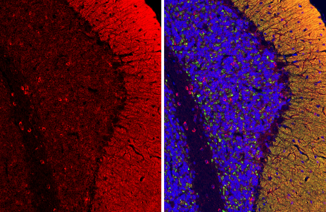
Tau antibody detects Tau protein by immunohistochemical analysis.Sample: Paraffin-embedded rat cerebellum.Red: Tau stained by Tau antibody (GTX130462) diluted at 1:250.Green: VGluT1 antibody [GT34] (GTX641142) diluted at 1:500.Blue: Hoechst 33342 staining.Antigen Retrieval: Citrate buffer, pH 6.0, 15 min
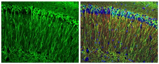
Tau antibody detects Tau protein expression by immunohistochemical analysis.
Sample: Frozen sectioned E13.5 Rat brain.
Green: Tau protein stained by Tau antibody (GTX130462) diluted at 1:250.
Red: beta Tubulin 3/ TUJ1, a mature neuron marker, stained by beta Tubulin 3/ TUJ1 antibody [GT11710] (GTX631836) diluted at 1:500.
Blue: Fluoroshield with DAPI (GTX30920).
Tau antibody detects Tau protein at cytoplasm by immunohistochemical analysis.Sample: Paraffin-embedded mouse brain.Green: Tau stained by Tau antibody (GTX130462) diluted at 1:250.Red: alpha Tubulin, a cytoskeleton marker, stained by alpha Tubulin antibody [GT114] (GTX628802) diluted at 1:1000.Blue: Fluoroshield with DAPI (GTX30920).Antigen Retrieval: Citrate buffer, pH 6.0, 15 min
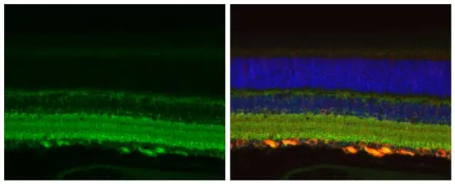
Tau antibody detects Tau protein at cytoplasm by immunohistochemical analysis.Sample: Paraffin-embedded mouse eye.Green: Tau stained by Tau antibody (GTX130462) diluted at 1:500.Red: beta Tubulin 3/ Tuj1 stained by beta Tubulin 3/ Tuj1 antibody [GT11710] (GTX631836) diluted at 1:500.Blue: Fluoroshield with DAPI (GTX30920).Antigen Retrieval: Citrate buffer, pH 6.0, 15 min
Rat tissue extract (50 μg) was separated by 7.5% SDS-PAGE, and the membrane was blotted with Tau antibody (GTX130462) diluted at 1:100000. The HRP-conjugated anti-rabbit IgG antibody (GTX213110-01) was used to detect the primary antibody.
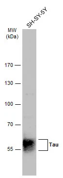
Whole cell extract (30 μg) was separated by 7.5% SDS-PAGE, and the membrane was blotted with Tau antibody (GTX130462) diluted at 1:1000. The HRP-conjugated anti-rabbit IgG antibody (GTX213110-01) was used to detect the primary antibody.
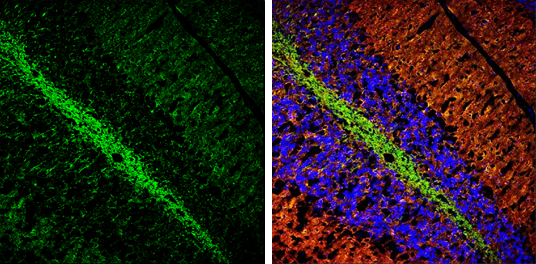
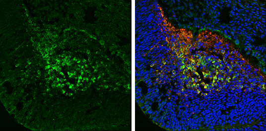
Tau antibody detects Tau protein expression by immunohistochemical analysis.
Sample: Frozen sectioned adult mouse cerebellum.
Green: Tau protein stained by Tau antibody (GTX130462) diluted at 1:250.
Red: beta Tubulin 3/ TUJ1, a stained by beta Tubulin 3/ TUJ1 antibody [GT11710] (GTX631836) diluted at 1:500.
Blue: Fluoroshield with DAPI (GTX30920).
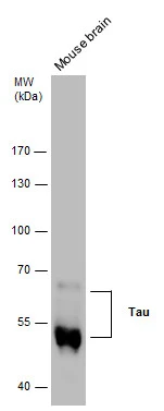
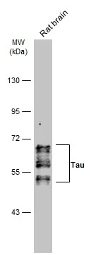
Tau antibody detects Tau protein by western blot analysis. Mouse tissue extracts (50 μg) was separated by 7.5 % SDS-PAGE, and the membrane was blotted with Tau antibody (GTX130462) at a dilution of 1:10000.
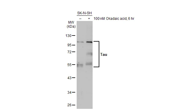
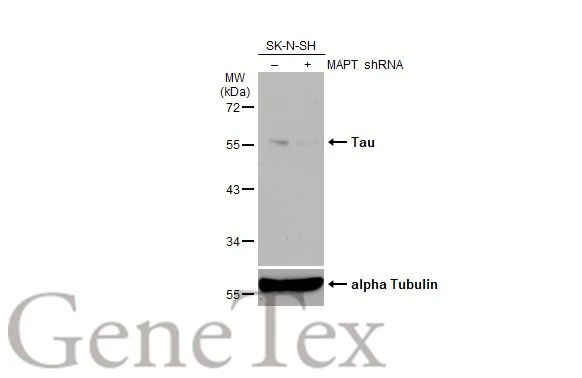
Untreated (–) and treated (+) SK-N-SH whole cell extract (30 μg) were separated by 10% SDS-PAGE, and the membrane was blotted with Tau antibody (GTX130462) diluted at 1:1000. The HRP-conjugated anti-rabbit IgG antibody (GTX213110-01) was used to detect the primary antibody, and the signal was developed with Trident ECL plus-Enhanced.
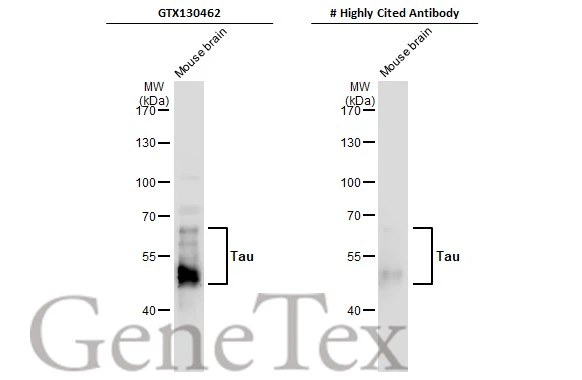
Mouse tissue extract (50 μg) was separated by 7.5% SDS-PAGE, and the membranes were blotted with Tau antibody (GTX130462) diluted at 1:4000 and competitor's antibody diluted at 1:2000. The HRP-conjugated anti-rabbit IgG antibody (GTX213110-01) was used to detect the primary antibody.
*The competitor is not affiliated with GeneTex and does not endorse this product.
-
宿主Rabbit
-
克隆Polyclonal
-
同种型IgG
-
实验应用WB ICC/IF IHC-P IHC-Fr
-
种属反应Human, Mouse, Rat