UBE2N抗体
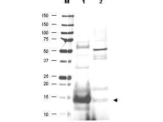
WB analysis of various samples using GTX48485 UBE2N antibody.
Lane 1 : Human small intestine tissue lysate
Lane 2 : Mouse thymus tissue lysate
Loading : 20 μg
Dilution : 1:500
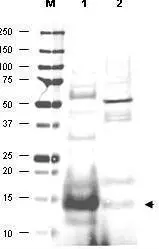
Western blot using GeneTex's affinity purified anti-UBC13 antibody shows detection of UBC13 protein in human small intestine lysate (lane 1) but not in mouse thymus lysate (lane 2). The heavily stained band in lane 1 (arrowhead) indicates this particular gel was overloaded with protein. The identity of minor reactive bands is unknown but could represent E2 complexes. Each lane contains approximately 20 μg of lysate. Primary antibody was used at a 1:500 dilution. The membrane was washed and reacted with a 1:10,000 dilution of Alexa FluorTM 680 conjugated rabbit anti-Goat IgG. Molecular weight estimation was made by comparison to prestained MW markers indicated at the left (lane M). Other detection systems will yield similar results.
-
宿主Goat
-
克隆Polyclonal
-
同种型IgG
-
实验应用WB ELISA
-
种属反应Human