PARP抗体


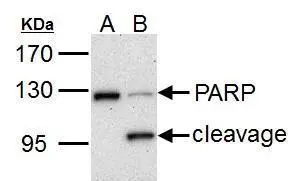
PARP1 antibody detects PARP1 protein by western blot analysis.
A. 30 μg HCT116 whole cell lysate/extract (untreated)
B. 30 μg HCT116 whole cell lysate/extract (30 μM cisplatin treatment for 24hr)
7.5% SDS-PAGE
PARP1 antibody (GTX100573) dilution: 1:1000
The HRP-conjugated anti-rabbit IgG antibody (GTX213110-01) was used to detect the primary antibody.
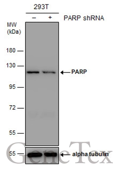
Non-transfected (–) and transfected (+) 293T whole cell extracts (30 μg) were separated by 7.5% SDS-PAGE, and the membrane was blotted with PARP antibody (GTX100573) diluted at 1:2000. The HRP-conjugated anti-rabbit IgG antibody (GTX213110-01) was used to detect the primary antibody.
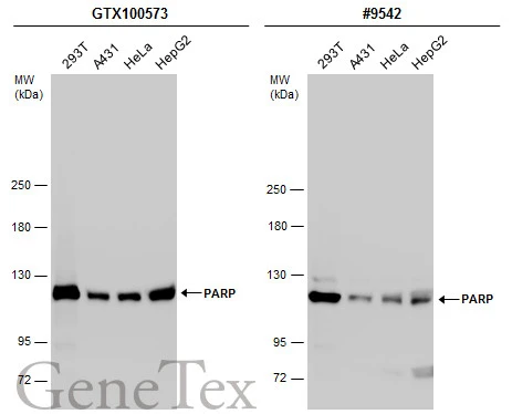
Various whole cell extracts (30 μg) were separated by 5% SDS-PAGE, and the membranes were blotted with PARP antibody (GTX100573) diluted at 1:2000 and competitor's antibody (#9542) diluted at 1:500. The HRP-conjugated anti-rabbit IgG antibody (GTX213110-01) was used to detect the primary antibody.
*The competitor is not affiliated with GeneTex and does not endorse this product.
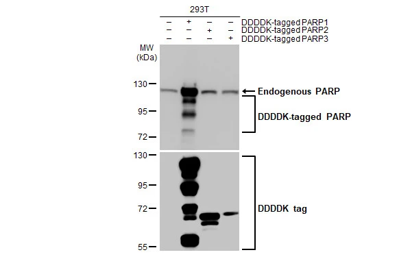
Non-transfected (–) and transfected (+) 293T whole cell extracts (30 μg) were separated by 7.5% SDS-PAGE, and the membrane was blotted with PARP antibody (GTX100573) diluted at 1:50000. The HRP-conjugated anti-rabbit IgG antibody (GTX213110-01) was used to detect the primary antibody.
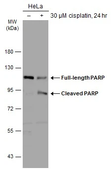
Untreated (–) and treated (+) HeLa whole cell extracts (30 μg) were separated by 7.5% SDS-PAGE, and the membrane was blotted with PARP antibody (GTX100573) diluted at 1:2000. The HRP-conjugated anti-rabbit IgG antibody (GTX213110-01) was used to detect the primary antibody.
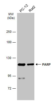
Various whole cell extracts (30 μg) were separated by 5% SDS-PAGE, and the membrane was blotted with PARP antibody (GTX100573) diluted at 1:1000. The HRP-conjugated anti-rabbit IgG antibody (GTX213110-01) was used to detect the primary antibody.
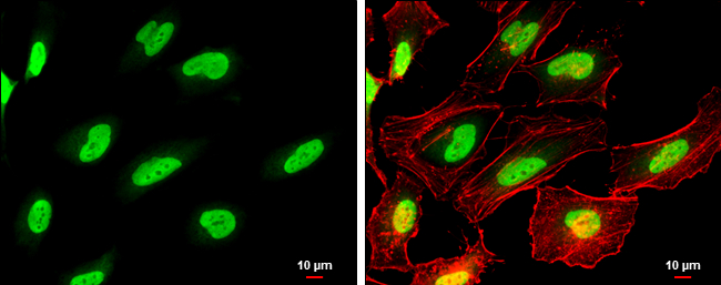
PARP antibody detects PARP protein at nucleus by immunofluorescent analysis.
Sample: HeLa cells were fixed in 4% paraformaldehyde at RT for 15 min.
Green: PARP protein stained by PARP antibody (GTX100573) diluted at 1:500.
Red: Phalloidin, a cytoskeleton marker, diluted at 1:100.
Scale bar = 10 μm.
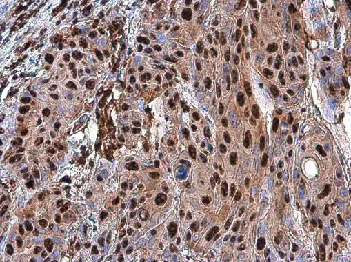
PARP antibody detects PARP protein at nucleus in human oral carcinoma by immunohistochemical analysis.
Sample: Paraffin-embedded human oral carcinoma.
PARP antibody (GTX100573) diluted at 1:500.
Antigen Retrieval: Citrate buffer, pH 6.0, 15 min
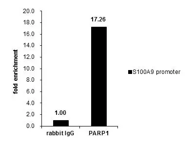
ChIP experiment and primer designs are based on BMC Mol Biol. 2006 Dec 22;7:48.
Cross-linked ChIP was performed with Raji chromatin extract and 5 μg of either control rabbit IgG or anti-PARP1 antibody. The precipitated DNA was detected by PCR with primer set targeting to S100A9 promoter.
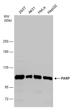
Various whole cell extracts (30 μg) were separated by 5% SDS-PAGE, and the membrane was blotted with PARP antibody (GTX100573) diluted at 1:2000.
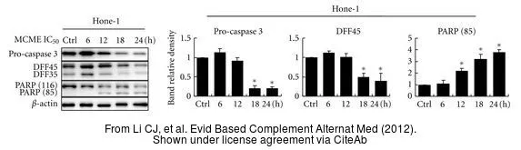
The data was published in the journal Evid Based Complement Alternat Med in 2012. PMID: 23091557
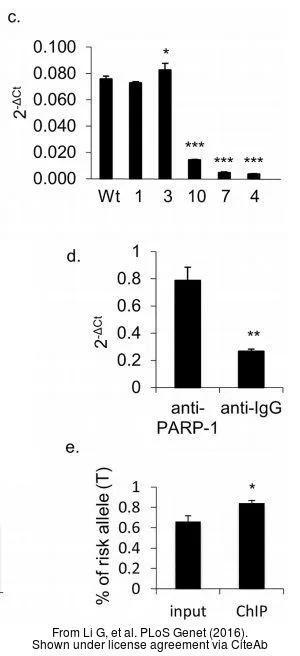
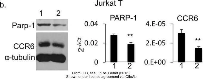
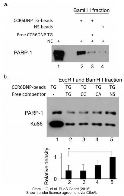
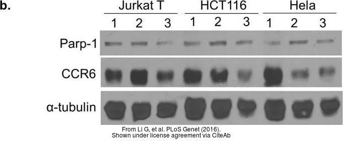
The data was published in the journal PLoS Genet in 2016. PMID: 27626929
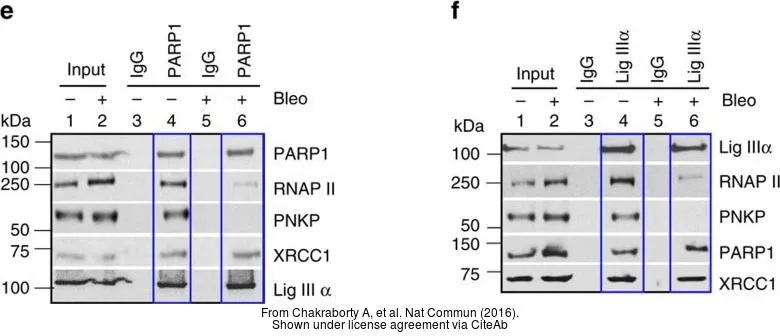
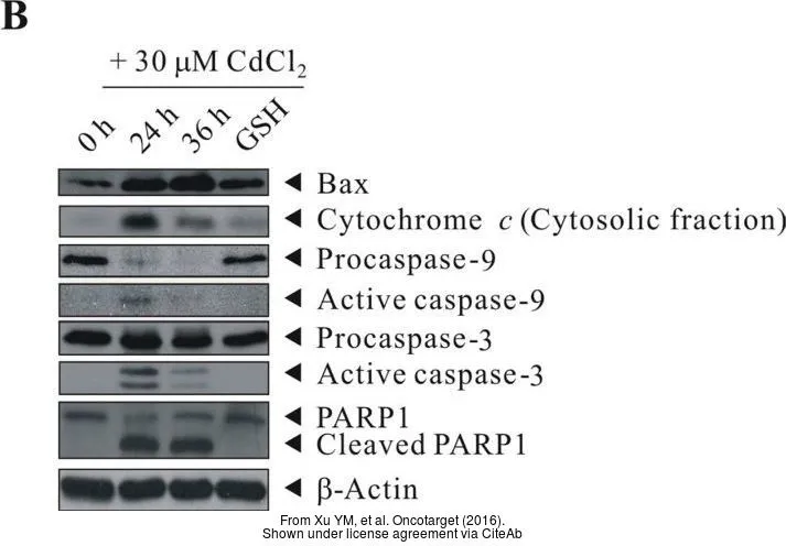
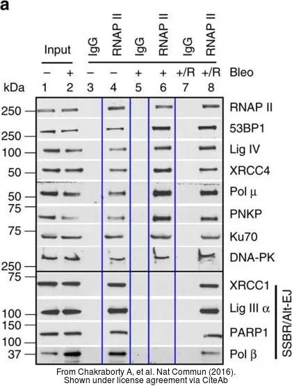
The data was published in the journal Nat Commun in 2016.PMID: 27703167
-
宿主Rabbit
-
克隆Polyclonal
-
同种型IgG
-
实验应用WB ICC/IF IHC-P IP ChIP assay
-
种属反应Human, Mouse, Rat