Mitochondrial dysfunction is a frequent cause of liver disease, with fission and fusion being key processes that maintain mitochondrial health. Mouse models with systemic knockouts (KO) of individual factors that mediate fission or fusion (such as Drp1, Mfn1, Mfn2, and OPA1) are all embryonic lethal, as are tissue-specific KOs in heart and brain. However, the published literature offers a more complex picture of the liver-specific function of OPA1 (optic atrophy 1), a fusion factor that is also involved in mitochondrial cristae structure. Some studies indicated that OPA1 depletion in the mouse liver imparts some protection from non-alcoholic fatty liver disease (NAFLD) and nonalcoholic steatohepatitis (NASH) pathologies.
To further explore the role of OPA1 in the liver, Lee et al. generated OPA1 liver knockout (LKO) mice (1). Instead of the expected severe metabolic impairment or outright lethality, the authors found that LKO mice are healthy, showing that OPA1 is dispensable in the liver. Though structural changes in the cristae were evident, mitochondrial respiration was unperturbed. Proteomic analyses indicated that the loss of OPA1 induced mitohormesis, a process where mild mitochondrial stress leads to a realigned homeostatic state in the cell that can protect it from additional insults. In this case, the authors found that mitohormesis resulted in almost complete resistance to liver toxicity caused by paracetamol/APAP exposure, a very common cause of severe hepatocyte death. This occurred through two mechanisms: reduced drug metabolism and elevated resistance to mitochondrial permeability transition. Thus, Lee et al. have identified a protective mitohormetic response in OPA1 LKO mice that supports liver resiliency.
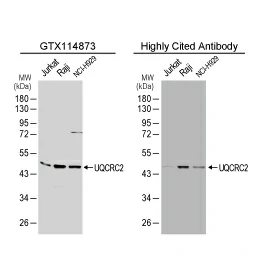
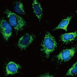
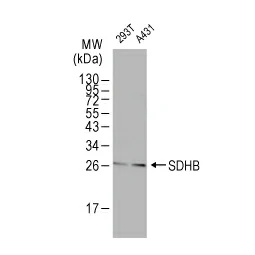
GeneTex offers an extensive catalog of quality antibodies and reagents for mitochondria and metabolism research, including the NDUFA5 antibody [N1C3] (GTX111016), SDHB antibody (GTX113833), UQCRC2 antibody (GTX114873), and COX4 antibody (GTX114330) cited in the Lee et al. study. In addition, GeneTex is excited to introduce its extensively validated recombinant rabbit monoclonal SDHB antibody [HL2251] (GTX638300) and COX4 antibody [HL2264] (GTX638316). For more information, please see the product data images below and visit www.genetex.com.
Highlighted Products
Reference:
- Nat Commun. 2023 Oct 23;14(1):6721. doi: 10.1038/s41467-023-42564-0.

![NDUFA5 antibody [N1C3] (GTX111016) NDUFA5 antibody [N1C3] (GTX111016)](/upload/media/MarketingMaterial/Newsletter/2023/OPA1/landingPage_img_255x255_01.webp)


![COX4 antibody [HL2264] (GTX638316) COX4 antibody [HL2264] (GTX638316)](/upload/media/MarketingMaterial/Newsletter/2023/OPA1/landingPage_img_255x255_04.webp)

![SDHB antibody [HL2251] (GTX638300) SDHB antibody [HL2251] (GTX638300)](/upload/media/MarketingMaterial/Newsletter/2023/OPA1/landingPage_img_255x255_06.webp)
![Cytochrome C antibody [HL2205] (GTX638209) Cytochrome C antibody [HL2205] (GTX638209)](/upload/media/MarketingMaterial/Newsletter/2023/OPA1/landingPage_img_255x255_07.webp)
![NRF2 antibody [HL1021] (GTX635826) NRF2 antibody [HL1021] (GTX635826)](/upload/media/MarketingMaterial/Newsletter/2023/OPA1/landingPage_img_255x255_08.webp)
![SOD2 antibody [HL1483] (GTX636957) SOD2 antibody [HL1483] (GTX636957)](/upload/media/MarketingMaterial/Newsletter/2023/OPA1/landingPage_img_255x255_09.webp)